 Movie
Movie Controller


+ Open data

- Basic information
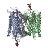
| Entry | Database: PDB / ID: 2ped | |||||||||
|---|---|---|---|---|---|---|---|---|---|---|
| Title | Crystallographic model of 9-cis-rhodopsin | |||||||||
 Components Components | Rhodopsin | |||||||||
 Keywords Keywords | SIGNALING PROTEIN / RHODOPSIN / G PROTEIN-COUPLED RECEPTOR / VISUAL PIGMENT | |||||||||
| Function / homology |  Function and homology informationOpsins / VxPx cargo-targeting to cilium / rod photoreceptor outer segment / rod bipolar cell differentiation / sperm head plasma membrane / podosome assembly / absorption of visible light / opsin binding / The canonical retinoid cycle in rods (twilight vision) / : ...Opsins / VxPx cargo-targeting to cilium / rod photoreceptor outer segment / rod bipolar cell differentiation / sperm head plasma membrane / podosome assembly / absorption of visible light / opsin binding / The canonical retinoid cycle in rods (twilight vision) / : / G protein-coupled photoreceptor activity / photoreceptor inner segment membrane / rhodopsin mediated signaling pathway / 11-cis retinal binding / cellular response to light stimulus / G protein-coupled receptor complex / Inactivation, recovery and regulation of the phototransduction cascade / phototransduction, visible light / thermotaxis / Activation of the phototransduction cascade / detection of temperature stimulus involved in thermoception / outer membrane / arrestin family protein binding / photoreceptor cell maintenance / photoreceptor outer segment membrane / G alpha (i) signalling events / response to light stimulus / phototransduction / photoreceptor outer segment / G-protein alpha-subunit binding / sperm midpiece / visual perception / guanyl-nucleotide exchange factor activity / microtubule cytoskeleton organization / photoreceptor disc membrane / cell-cell junction / gene expression / G protein-coupled receptor signaling pathway / Golgi membrane / zinc ion binding / membrane / identical protein binding / plasma membrane Function and homology informationOpsins / VxPx cargo-targeting to cilium / rod photoreceptor outer segment / rod bipolar cell differentiation / sperm head plasma membrane / podosome assembly / absorption of visible light / opsin binding / The canonical retinoid cycle in rods (twilight vision) / : ...Opsins / VxPx cargo-targeting to cilium / rod photoreceptor outer segment / rod bipolar cell differentiation / sperm head plasma membrane / podosome assembly / absorption of visible light / opsin binding / The canonical retinoid cycle in rods (twilight vision) / : / G protein-coupled photoreceptor activity / photoreceptor inner segment membrane / rhodopsin mediated signaling pathway / 11-cis retinal binding / cellular response to light stimulus / G protein-coupled receptor complex / Inactivation, recovery and regulation of the phototransduction cascade / phototransduction, visible light / thermotaxis / Activation of the phototransduction cascade / detection of temperature stimulus involved in thermoception / outer membrane / arrestin family protein binding / photoreceptor cell maintenance / photoreceptor outer segment membrane / G alpha (i) signalling events / response to light stimulus / phototransduction / photoreceptor outer segment / G-protein alpha-subunit binding / sperm midpiece / visual perception / guanyl-nucleotide exchange factor activity / microtubule cytoskeleton organization / photoreceptor disc membrane / cell-cell junction / gene expression / G protein-coupled receptor signaling pathway / Golgi membrane / zinc ion binding / membrane / identical protein binding / plasma membraneSimilarity search - Function | |||||||||
| Biological species |  Bos taurus (cattle) Bos taurus (cattle) | |||||||||
| Method | X-RAY DIFFRACTION / SYNCHROTRON / MOLECULAR REPLACEMENT / Resolution: 2.95 Å | |||||||||
 Authors Authors | Nakamichi, H. / Okada, T. | |||||||||
 Citation Citation | Journal: Biophys.J. / Year: 2007 Title: Photoisomerization mechanism of rhodopsin and 9-cis-rhodopsin revealed by x-ray crystallography Authors: Nakamichi, H. / Buss, V. / Okada, T. #1: Journal: J.Am.Chem.Soc. / Year: 2007 Title: Protein assistance in the photoisomerization of rhodopsin and 9-cis-rhodopsin--insights from experiment and theory Authors: Sekharan, S. / Sugihara, M. / Weingart, O. / Okada, T. / Buss, V. | |||||||||
| History |
|
- Structure visualization
| Structure viewer | Molecule: MolmilJmol/JSmol |
|---|
- Downloads & links
-Download
| PDBx/mmCIF format | 2ped.cif.gz | 163.6 KB | Display | PDBx/mmCIF format |
|---|---|---|---|---|
| PDB format | pdb2ped.ent.gz | 128.3 KB | Display | PDB format |
| PDBx/mmJSON format | 2ped.json.gz | Tree view | PDBx/mmJSON format | |
| Others |  Other downloads Other downloads |
-Validation report
| Arichive directory | https://data.pdbj.org/pub/pdb/validation_reports/pe/2pedftp://data.pdbj.org/pub/pdb/validation_reports/pe/2ped | HTTPS FTP |
|---|
-Related structure data
| Related structure data | 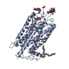 1u19S S: Starting model for refinement |
|---|---|
| Similar structure data |


-Links
 PDBj
PDBj


- Assembly
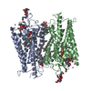
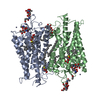
| Deposited unit | 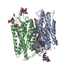
| ||||||||
|---|---|---|---|---|---|---|---|---|---|
| 1 |
| ||||||||
| 2 | 
| ||||||||
| Unit cell |
|
-Components
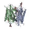
-Protein , 1 types, 2 molecules AB
| #1: Protein | Mass: 39057.492 Da / Num. of mol.: 2 / Source method: isolated from a natural source / Source: (natural) Bos taurus (cattle) / References: UniProt: P02699 |
|---|
-Sugars , 4 types, 8 molecules 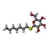
| #2: Polysaccharide | alpha-D-mannopyranose-(1-4)-2-acetamido-2-deoxy-beta-D-glucopyranose-(1-4)-2-acetamido-2-deoxy-beta- ...alpha-D-mannopyranose-(1-4)-2-acetamido-2-deoxy-beta-D-glucopyranose-(1-4)-2-acetamido-2-deoxy-beta-D-glucopyranose / Mass: 586.542 Da / Num. of mol.: 1 Source method: isolated from a genetically manipulated source | ||||
|---|---|---|---|---|---|
| #3: Polysaccharide | / Mass: 424.401 Da / Num. of mol.: 2 Source method: isolated from a genetically manipulated source #4: Polysaccharide | beta-D-mannopyranose-(1-3)-beta-D-mannopyranose-(1-4)-2-acetamido-2-deoxy-beta-D-glucopyranose-(1-4) ...beta-D-mannopyranose-(1-3)-beta-D-mannopyranose-(1-4)-2-acetamido-2-deoxy-beta-D-glucopyranose-(1-4)-2-acetamido-2-deoxy-beta-D-glucopyranose | / Mass: 748.682 Da / Num. of mol.: 1Source method: isolated from a genetically manipulated source #9: Sugar | ChemComp-HTG /  Type: D-saccharide / Mass: 294.408 Da / Num. of mol.: 4 / Source method: obtained synthetically / Formula: C13H26O5S / Comment: detergent*YM Type: D-saccharide / Mass: 294.408 Da / Num. of mol.: 4 / Source method: obtained synthetically / Formula: C13H26O5S / Comment: detergent*YM |
-Non-polymers , 6 types, 130 molecules 


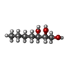

| #5: Chemical | ChemComp-HG / Mercury (element) Mass: 200.590 Da / Num. of mol.: 6 / Source method: obtained synthetically / Formula: Hg Mass: 200.590 Da / Num. of mol.: 6 / Source method: obtained synthetically / Formula: Hg#6: Chemical | ChemComp-ZN /  Mass: 65.409 Da / Num. of mol.: 7 / Source method: obtained synthetically / Formula: Zn Mass: 65.409 Da / Num. of mol.: 7 / Source method: obtained synthetically / Formula: Zn#7: Chemical | Retinal Mass: 284.436 Da / Num. of mol.: 2 / Source method: obtained synthetically / Formula: C20H28O Mass: 284.436 Da / Num. of mol.: 2 / Source method: obtained synthetically / Formula: C20H28O#8: Chemical | ChemComp-PLM / Palmitic acid Mass: 256.424 Da / Num. of mol.: 6 / Source method: obtained synthetically / Formula: C16H32O2 Mass: 256.424 Da / Num. of mol.: 6 / Source method: obtained synthetically / Formula: C16H32O2#10: Chemical | ChemComp-HTO / |  Mass: 148.200 Da / Num. of mol.: 1 / Source method: obtained synthetically / Formula: C7H16O3 Mass: 148.200 Da / Num. of mol.: 1 / Source method: obtained synthetically / Formula: C7H16O3#11: Water | ChemComp-HOH / | WaterMass: 18.015 Da / Num. of mol.: 108 / Source method: isolated from a natural source / Formula: H2O |
|---|
-Experimental details
-Experiment
| Experiment | Method: X-RAY DIFFRACTION / Number of used crystals: 1 |
|---|
- Sample preparation
| Crystal | Density Matthews: 4.44 Å3/Da / Density % sol: 72.32 % |
|---|---|
| Crystal grow | Temperature: 283 K / Method: vapor diffusion, hanging drop / pH: 6 Details: 2.9M AMMONIUM SULFATE, 0.05M MES, pH 6.0, VAPOR DIFFUSION, HANGING DROP, temperature 283K |
-Data collection
| Diffraction | Mean temperature: 100 K |
|---|---|
| Diffraction source | Source: SYNCHROTRON / Site: SPring-8  / Beamline: BL41XU / Wavelength: 1 Å / Beamline: BL41XU / Wavelength: 1 Å |
| Detector | Type: MAR CCD 165 mm / Detector: CCD / Date: Sep 25, 2003 |
| Radiation | Protocol: SINGLE WAVELENGTH / Monochromatic (M) / Laue (L): M / Scattering type: x-ray |
| Radiation wavelength | Wavelength: 1 Å / Relative weight: 1 |
| Reflection | Resolution: 2.9→50 Å / Num. obs: 27964 / % possible obs: 91.8 % / Observed criterion σ(I): 0 / Redundancy: 3.5 % / Biso Wilson estimate: 56.8 Å2 / Rmerge(I) obs: 0.139 / Net I/σ(I): 8.27 |
| Reflection shell | Resolution: 2.9→3 Å / Redundancy: 2.5 % / Rmerge(I) obs: 0.778 / Mean I/σ(I) obs: 1.06 / Num. unique all: 1615 / % possible all: 53.2 |
- Processing
| Software |
| |||||||||||||||||||||||||
|---|---|---|---|---|---|---|---|---|---|---|---|---|---|---|---|---|---|---|---|---|---|---|---|---|---|---|
| Refinement | Method to determine structure: MOLECULAR REPLACEMENT Starting model: PDB ENTRY 1U19 Resolution: 2.95→50 Å / Cross valid method: THROUGHOUT / σ(F): 0
| |||||||||||||||||||||||||
| Displacement parameters | Biso mean: 76.849 Å2 | |||||||||||||||||||||||||
| Refine analyze |
| |||||||||||||||||||||||||
| Refinement step | Cycle: LAST / Resolution: 2.95→50 Å
| |||||||||||||||||||||||||
| Refine LS restraints |
|
