 Movie
Movie Controller

[English] 日本語
 Yorodumi
- PDB-2pe7: Thaumatin from Thaumatococcus Danielli in complex with tris-dipic... -
+ Open data

- Basic information
| Entry | Database: PDB / ID: 2pe7 | ||||||
|---|---|---|---|---|---|---|---|
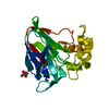
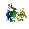
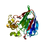
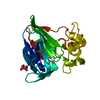
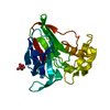
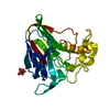
| Title | Thaumatin from Thaumatococcus Danielli in complex with tris-dipicolinate Europium | ||||||
 Components Components | Preprothaumatin I | ||||||
 Keywords Keywords |  PLANT PROTEIN / Thaumatin / Tris-dipicolinate Europium PLANT PROTEIN / Thaumatin / Tris-dipicolinate Europium | ||||||
| Function / homology |  Function and homology information Function and homology information | ||||||
| Biological species |  Thaumatococcus daniellii (katemfe) Thaumatococcus daniellii (katemfe) | ||||||
| Method | X-RAY DIFFRACTION / SYNCHROTRON / MAD / Resolution: 1.46 Å | ||||||
 Authors Authors | Pompidor, G. / Vicat, J. / Kahn, R. | ||||||
 Citation Citation | Journal: Acta Crystallogr.,Sect.D / Year: 2010 Title: A dipicolinate lanthanide complex for solving protein structures using anomalous diffraction. Authors: Pompidor, G. / Maury, O. / Vicat, J. / Kahn, R. | ||||||
| History |
|
- Structure visualization
| Structure viewer | Molecule: MolmilJmol/JSmol |
|---|
- Downloads & links
-Download
| PDBx/mmCIF format | 2pe7.cif.gz | 108.6 KB | Display | PDBx/mmCIF format |
|---|---|---|---|---|
| PDB format | pdb2pe7.ent.gz | 81.8 KB | Display | PDB format |
| PDBx/mmJSON format | 2pe7.json.gz | Tree view | PDBx/mmJSON format | |
| Others |  Other downloads Other downloads |
-Validation report
| Arichive directory | https://data.pdbj.org/pub/pdb/validation_reports/pe/2pe7ftp://data.pdbj.org/pub/pdb/validation_reports/pe/2pe7 | HTTPS FTP |
|---|
-Related structure data
| Related structure data |  2pesC  3lgrC 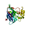 1rqwS S: Starting model for refinement C: citing same article ( |
|---|---|
| Similar structure data |


-Links
 PDBj
PDBj

- Assembly
| Deposited unit | 
| ||||||||||||
|---|---|---|---|---|---|---|---|---|---|---|---|---|---|
| 1 |
| ||||||||||||
| Unit cell |
| ||||||||||||
| Components on special symmetry positions |
|
-Components
| #1: Protein | Mass: 22228.043 Da / Num. of mol.: 1 / Source method: isolated from a natural source / Source: (natural) Thaumatococcus daniellii (katemfe) / References: UniProt: A1IIJ1, UniProt: P02883*PLUS | ||||||||
|---|---|---|---|---|---|---|---|---|---|
| #2: Chemical | ChemComp-EU / Europium  Mass: 151.964 Da / Num. of mol.: 1 / Source method: obtained synthetically / Formula: Eu Mass: 151.964 Da / Num. of mol.: 1 / Source method: obtained synthetically / Formula: Eu | ||||||||
| #3: Chemical | Tartaric acid  Mass: 150.087 Da / Num. of mol.: 2 / Source method: obtained synthetically / Formula: C4H6O6 Mass: 150.087 Da / Num. of mol.: 2 / Source method: obtained synthetically / Formula: C4H6O6#4: Chemical | Dipicolinic acid  Mass: 167.119 Da / Num. of mol.: 3 / Source method: obtained synthetically / Formula: C7H5NO4 Mass: 167.119 Da / Num. of mol.: 3 / Source method: obtained synthetically / Formula: C7H5NO4#5: Water | ChemComp-HOH / | Water Mass: 18.015 Da / Num. of mol.: 351 / Source method: isolated from a natural source / Formula: H2O Mass: 18.015 Da / Num. of mol.: 351 / Source method: isolated from a natural source / Formula: H2ONonpolymer details | ACCORDING TO AUTHORS THE LIGAND PDC IS IN THE BASIC (DEPROTONAT | Sequence details | THERE ARE TWO ISOFORMS OF THAUMATIN, I AND II. CRYSTALLIZATION MATERIAL CONTAINS A MIXTURE OF LYS46 ...THERE ARE TWO ISOFORMS OF THAUMATIN, I AND II. CRYSTALLIZ | |
-Experimental details
-Experiment
| Experiment | Method: X-RAY DIFFRACTION / Number of used crystals: 1 |
|---|
- Sample preparation
| Crystal | Density Matthews: 2.78 Å3/Da / Density % sol: 55.74 % |
|---|---|
| Crystal grow | Temperature: 293 K / Method: vapor diffusion, hanging drop / pH: 6.5 Details: 0.7 M Sodium Potassium Tatrate, pH 6.5, VAPOR DIFFUSION, HANGING DROP, temperature 293K |
-Data collection
| Diffraction |
| ||||||||||||||||||||||||||||||||||||||||||||||||||||||||||||||||||||||||||||||||||||||||
|---|---|---|---|---|---|---|---|---|---|---|---|---|---|---|---|---|---|---|---|---|---|---|---|---|---|---|---|---|---|---|---|---|---|---|---|---|---|---|---|---|---|---|---|---|---|---|---|---|---|---|---|---|---|---|---|---|---|---|---|---|---|---|---|---|---|---|---|---|---|---|---|---|---|---|---|---|---|---|---|---|---|---|---|---|---|---|---|---|---|
| Diffraction source |
| ||||||||||||||||||||||||||||||||||||||||||||||||||||||||||||||||||||||||||||||||||||||||
| Detector | Type: MAR CCD 165 mm / Detector: CCD / Date: Dec 25, 2004 / Details: Si(111) | ||||||||||||||||||||||||||||||||||||||||||||||||||||||||||||||||||||||||||||||||||||||||
| Radiation | Protocol: MAD / Monochromatic (M) / Laue (L): M / Scattering type: x-ray | ||||||||||||||||||||||||||||||||||||||||||||||||||||||||||||||||||||||||||||||||||||||||
| Radiation wavelength |
| ||||||||||||||||||||||||||||||||||||||||||||||||||||||||||||||||||||||||||||||||||||||||
| Reflection | Resolution: 1.456→45.596 Å / Num. obs: 44812 / % possible obs: 99.5 % / Observed criterion σ(I): 0 / Redundancy: 12.3 % / Biso Wilson estimate: 11.4 Å2 / Rmerge(I) obs: 0.074 / Rsym value: 0.074 / Net I/σ(I): 5.5 | ||||||||||||||||||||||||||||||||||||||||||||||||||||||||||||||||||||||||||||||||||||||||
| Reflection shell | Diffraction-ID: 1
|

-Phasing
| Phasing | Method: MAD | |||||||||||||||||||||||||||||||||||||||||||||||||||||||||||||||||||||||||||||||||||||||||||
|---|---|---|---|---|---|---|---|---|---|---|---|---|---|---|---|---|---|---|---|---|---|---|---|---|---|---|---|---|---|---|---|---|---|---|---|---|---|---|---|---|---|---|---|---|---|---|---|---|---|---|---|---|---|---|---|---|---|---|---|---|---|---|---|---|---|---|---|---|---|---|---|---|---|---|---|---|---|---|---|---|---|---|---|---|---|---|---|---|---|---|---|---|
| Phasing MAD set site |
|
- Processing
| Software |
| ||||||||||||||||||||||||||||||||||||||||||||||||||||||||||||||||||||||||||||||||||||||||||||||||||||||||||||||||||||||||||||||||||||||||||||
|---|---|---|---|---|---|---|---|---|---|---|---|---|---|---|---|---|---|---|---|---|---|---|---|---|---|---|---|---|---|---|---|---|---|---|---|---|---|---|---|---|---|---|---|---|---|---|---|---|---|---|---|---|---|---|---|---|---|---|---|---|---|---|---|---|---|---|---|---|---|---|---|---|---|---|---|---|---|---|---|---|---|---|---|---|---|---|---|---|---|---|---|---|---|---|---|---|---|---|---|---|---|---|---|---|---|---|---|---|---|---|---|---|---|---|---|---|---|---|---|---|---|---|---|---|---|---|---|---|---|---|---|---|---|---|---|---|---|---|---|---|---|
| Refinement | Method to determine structure: MAD Starting model: 1RQW Resolution: 1.46→53.68 Å / Cor.coef. Fo:Fc: 0.965 / Cor.coef. Fo:Fc free: 0.956 / Cross valid method: THROUGHOUT / σ(F): 0 / ESU R: 0.065 / ESU R Free: 0.057 / Stereochemistry target values: MAXIMUM LIKELIHOOD / Details: HYDROGENS HAVE BEEN ADDED IN THE RIDING POSITIONS
| ||||||||||||||||||||||||||||||||||||||||||||||||||||||||||||||||||||||||||||||||||||||||||||||||||||||||||||||||||||||||||||||||||||||||||||
| Solvent computation | Ion probe radii: 0.8 Å / Shrinkage radii: 0.8 Å / VDW probe radii: 1.2 Å / Solvent model: MASK | ||||||||||||||||||||||||||||||||||||||||||||||||||||||||||||||||||||||||||||||||||||||||||||||||||||||||||||||||||||||||||||||||||||||||||||
| Displacement parameters | Biso mean: 10.078 Å2
| ||||||||||||||||||||||||||||||||||||||||||||||||||||||||||||||||||||||||||||||||||||||||||||||||||||||||||||||||||||||||||||||||||||||||||||
| Refinement step | Cycle: LAST / Resolution: 1.46→53.68 Å
| ||||||||||||||||||||||||||||||||||||||||||||||||||||||||||||||||||||||||||||||||||||||||||||||||||||||||||||||||||||||||||||||||||||||||||||
| Refine LS restraints |
| ||||||||||||||||||||||||||||||||||||||||||||||||||||||||||||||||||||||||||||||||||||||||||||||||||||||||||||||||||||||||||||||||||||||||||||
| LS refinement shell | Resolution: 1.456→1.494 Å / Total num. of bins used: 20
|
