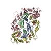
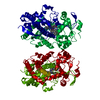
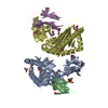
- PDB-2p6a: The structure of the Activin:Follistatin 315 complex -
+
Open data
ID or keywords:
Loading...
-
Basic information
Entry
Database: PDB / ID: 2p6a
Title
The structure of the Activin:Follistatin 315 complex
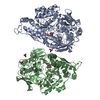
Components
Follistatin
Inhibin beta A chain
probable fragment of follistatin
Keywords
SIGNALING PROTEIN / Follistatin / Activin / Inhibin / TGF-beta
Function / homology
Function and homology information
activin receptor antagonist activity / activin A complex / inhibin A complex / cardiac fibroblast cell development / regulation of follicle-stimulating hormone secretion / negative regulation of B cell differentiation / positive regulation of ovulation / GABAergic neuron differentiation / Antagonism of Activin by Follistatin / negative regulation of follicle-stimulating hormone secretion ...activin receptor antagonist activity / activin A complex / inhibin A complex / cardiac fibroblast cell development / regulation of follicle-stimulating hormone secretion / negative regulation of B cell differentiation / positive regulation of ovulation / GABAergic neuron differentiation / Antagonism of Activin by Follistatin / negative regulation of follicle-stimulating hormone secretion / progesterone secretion / type II activin receptor binding / striatal medium spiny neuron differentiation / ameloblast differentiation / negative regulation of macrophage differentiation / Glycoprotein hormones / positive regulation of follicle-stimulating hormone secretion / cellular response to oxygen-glucose deprivation / hemoglobin biosynthetic process / positive regulation of hair follicle development / regulation of BMP signaling pathway / gamete generation / cellular response to follicle-stimulating hormone stimulus / cellular response to cholesterol / pattern specification process / Signaling by BMP / activin binding / negative regulation of phosphorylation / activin receptor signaling pathway / heparan sulfate proteoglycan binding / Signaling by Activin / negative regulation of activin receptor signaling pathway / mesodermal cell differentiation / positive regulation of extrinsic apoptotic signaling pathway in absence of ligand / SMAD protein signal transduction / cellular response to angiotensin / positive regulation of transcription by RNA polymerase III / odontogenesis / negative regulation of epithelial cell differentiation / response to aldosterone / hair follicle morphogenesis / negative regulation of G1/S transition of mitotic cell cycle / female gonad development / roof of mouth development / eyelid development in camera-type eye / endodermal cell differentiation / odontogenesis of dentin-containing tooth / peptide hormone binding / positive regulation of SMAD protein signal transduction / negative regulation of type II interferon production / keratinocyte proliferation / hair follicle development / positive regulation of collagen biosynthetic process / BMP signaling pathway / hematopoietic progenitor cell differentiation / extrinsic apoptotic signaling pathway / ovarian follicle development / positive regulation of protein metabolic process / erythrocyte differentiation / positive regulation of erythrocyte differentiation / skeletal system development / cytokine activity / growth factor activity / defense response / hormone activity / negative regulation of cell growth / response to organic cyclic compound / autophagy / cytokine-mediated signaling pathway / male gonad development / cell-cell signaling / nervous system development / cellular response to hypoxia / transcription by RNA polymerase II / cell differentiation / positive regulation of ERK1 and ERK2 cascade / cell surface receptor signaling pathway / positive regulation of protein phosphorylation / negative regulation of cell population proliferation / protein-containing complex binding / positive regulation of gene expression / regulation of transcription by RNA polymerase II / perinuclear region of cytoplasm / positive regulation of DNA-templated transcription / negative regulation of transcription by RNA polymerase II / positive regulation of transcription by RNA polymerase II / extracellular space / extracellular region / identical protein binding / nucleus / cytoplasm Similarity search - Function
Component-ID: 1 / Ens-ID: 1 / Beg auth comp-ID: GLY / Beg label comp-ID: GLY / End auth comp-ID: SER / End label comp-ID: SER / Refine code: 5 / Auth seq-ID: 1 - 116 / Label seq-ID: 1 - 116
Dom-ID
Auth asym-ID
Label asym-ID
1
A
A
2
B
C
Details
The biological assembly of these proteins is one activin A dimer in complex with 2 follistatin 315 molecules, which compose the activin:follistatin 315 complex. We observe one complex in the asymmetric unit.
Mass: 12991.865 Da / Num. of mol.: 2 Source method: isolated from a genetically manipulated source Source: (gene. exp.) Homo sapiens (human) / Gene: INHBA / Production host: Cricetulus griseus (Chinese hamster) / References: UniProt: P08476
#2: Protein
Follistatin / / FS / Activin-binding protein
Mass: 34796.379 Da / Num. of mol.: 2 Source method: isolated from a genetically manipulated source Source: (gene. exp.) Homo sapiens (human) / Gene: FST / Production host: Cricetulus griseus (Chinese hamster) / References: UniProt: P19883
#3: Protein/peptide
probablefragmentoffollistatin
Mass: 728.793 Da / Num. of mol.: 1 Source method: isolated from a genetically manipulated source
Sequence details
AUTHOR STATE THE FOLLOWING IN THE PUBLICATION: ON ONE FOLLISTATIN MOLECULE (CHAIN C AND ITS ...AUTHOR STATE THE FOLLOWING IN THE PUBLICATION: ON ONE FOLLISTATIN MOLECULE (CHAIN C AND ITS SYMMETRICALLY EQUIVALENT MOLECULES), ELECTRON DENSITY COULD BE SEEN FOR RESIDUES 289-299. ON THE SECOND FOLLISTATIN MOLECULE(CHAIN D AND ITS SYMMETRICALLY EQUIVALENT MOLECULES), CONTINUOUS ELECTRON DENSITY ALLOWED FOR THE BUILDING OF MAIN-CHAIN ATOMS OF 10 RESIDUES. SIDE CHAINS FOR THESE RESIDUES COULD NOT BE UNAMBIGUOUSLY MODELED, PREVENTING IDENTIFICATION OF THIS SEQUENCE IN THE C-TERMINAL EXTENSION. HOWEVER, BASED ON THE SYMMETRY OF THE TWO FOLLISTATIN MOLECULES, THIS STRETCH LIKELY CONSISTS OF RESIDUES 295-304, REPRESENTING THE MAJORITY OF THE CHARGED AMINO ACIDS IN THE C-TERMINAL EXTENSION.
-
Experimental details
-
Experiment
Experiment
Method: X-RAY DIFFRACTION / Number of used crystals: 1
-
Sample preparation
Crystal
Density Matthews: 2.53 Å3/Da / Density % sol: 51.47 %
Crystal grow
Temperature: 298 K / Method: vapor diffusion, hanging drop / pH: 6.5 Details: 20-23% PEG 1000, 200mM MgCl2, 3% EtOH, 20mM Trimethyl-amine HCl, pH 6.5, VAPOR DIFFUSION, HANGING DROP, temperature 298 K
Type: MARMOSAIC 300 mm CCD / Detector: CCD / Date: Jun 26, 2006
Radiation
Protocol: SINGLE WAVELENGTH / Monochromatic (M) / Laue (L): M / Scattering type: x-ray
Radiation wavelength
Wavelength: 1 Å / Relative weight: 1
Reflection
Redundancy: 7.2 % / Av σ(I) over netI: 4 / Number: 100464 / Rmerge(I) obs: 0.16 / Rsym value: 0.16 / D res high: 3.4 Å / D res low: 34.344 Å / Num. obs: 13978 / % possible obs: 99.7
Method: Solvent flattening and Histogram matching / Reflection: 13943
Phasing dm shell
Resolution (Å)
Delta phi final
FOM
Reflection
10.44-100
37.9
0.768
501
8.29-10.44
40.2
0.87
503
7.23-8.29
45.5
0.844
505
6.57-7.23
45.4
0.838
502
6.09-6.57
43
0.844
506
5.72-6.09
46.2
0.842
510
5.43-5.72
41.7
0.863
502
5.19-5.43
36.2
0.881
503
4.98-5.19
37.3
0.891
506
4.8-4.98
34.9
0.883
513
4.65-4.8
36.1
0.885
516
4.51-4.65
32.3
0.9
539
4.38-4.51
34.2
0.888
532
4.26-4.38
34.2
0.897
552
4.15-4.26
36.2
0.9
575
4.05-4.15
36.8
0.888
581
3.95-4.05
32.5
0.885
605
3.87-3.95
36.4
0.877
620
3.78-3.87
39.6
0.861
618
3.71-3.78
33.6
0.881
643
3.63-3.71
36.1
0.875
650
3.56-3.63
43.3
0.833
663
3.5-3.56
40.8
0.845
658
3.4-3.5
42.9
0.834
1140
-
Processing
Software
Name
Version
Classification
NB
SCALA
datascaling
PHASER
phasing
DM
phasing
REFMAC
refinement
PDB_EXTRACT
2
dataextraction
MOSFLM
datareduction
CCP4
(SCALA)
datascaling
Refinement
Method to determine structure: MOLECULAR REPLACEMENT Starting model: one activin A monomer and ND, FSD1, and FSD2 of one follistatin 288 molecule (PDB 2B0U)
Resolution: 3.4→31.01 Å / Cor.coef. Fo:Fc: 0.913 / Cor.coef. Fo:Fc free: 0.811 / SU B: 90.513 / SU ML: 0.667 / Cross valid method: THROUGHOUT / σ(F): 0 / ESU R Free: 0.733 / Stereochemistry target values: MAXIMUM LIKELIHOOD / Details: HYDROGENS HAVE BEEN ADDED IN THE RIDING POSITIONS
Rfactor
Num. reflection
% reflection
Selection details
Rfree
0.324
283
2 %
RANDOM
Rwork
0.223
-
-
-
obs
0.225
13955
99.69 %
-
Solvent computation
Ion probe radii: 0.8 Å / Shrinkage radii: 0.8 Å / VDW probe radii: 1.4 Å / Solvent model: MASK
In the structure databanks used in Yorodumi, some data are registered as the other names, "COVID-19 virus" and "2019-nCoV". Here are the details of the virus and the list of structure data.
Jan 31, 2019. EMDB accession codes are about to change! (news from PDBe EMDB page)
EMDB accession codes are about to change! (news from PDBe EMDB page)
The allocation of 4 digits for EMDB accession codes will soon come to an end. Whilst these codes will remain in use, new EMDB accession codes will include an additional digit and will expand incrementally as the available range of codes is exhausted. The current 4-digit format prefixed with “EMD-” (i.e. EMD-XXXX) will advance to a 5-digit format (i.e. EMD-XXXXX), and so on. It is currently estimated that the 4-digit codes will be depleted around Spring 2019, at which point the 5-digit format will come into force.
The EM Navigator/Yorodumi systems omit the EMD- prefix.
Related info.:Q: What is EMD? / ID/Accession-code notation in Yorodumi/EM Navigator
Yorodumi is a browser for structure data from EMDB, PDB, SASBDB, etc.
This page is also the successor to EM Navigator detail page, and also detail information page/front-end page for Omokage search.
The word "yorodu" (or yorozu) is an old Japanese word meaning "ten thousand". "mi" (miru) is to see.
Related info.:EMDB / PDB / SASBDB / Comparison of 3 databanks / Yorodumi Search / Aug 31, 2016. New EM Navigator & Yorodumi / Yorodumi Papers / Jmol/JSmol / Function and homology information / Changes in new EM Navigator and Yorodumi
 Movie
Movie Controller


 Open data
 Basic information
 Components
 Keywords
 SIGNALING PROTEIN /
  Function and homology information
 Authors
 Citation
 Structure visualization
 Downloads & links
 Other downloads


 PDBj
PDBj


 Assembly
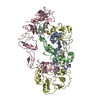


 Sample preparation
 / Beamline: 5ID-B / Wavelength: 1 Å
 Processing