 Movie
Movie Controller

[English] 日本語
 Yorodumi
- PDB-2p5t: Molecular and structural characterization of the PezAT chromosoma... -
+ Open data

- Basic information
| Entry | Database: PDB / ID: 2p5t | ||||||
|---|---|---|---|---|---|---|---|
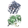
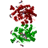
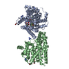
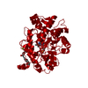
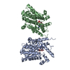
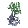
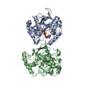
| Title | Molecular and structural characterization of the PezAT chromosomal toxin-antitoxin system of the human pathogen Streptococcus pneumoniae | ||||||
 Components Components |
| ||||||
 Keywords Keywords |  TRANSCRIPTION REGULATOR / postsegregational killing system / phosphoryltransferase / helix-turn-helix motif TRANSCRIPTION REGULATOR / postsegregational killing system / phosphoryltransferase / helix-turn-helix motif | ||||||
| Function / homology |  Function and homology informationUDP-N-acetylglucosamine kinase / : / kinase activity / DNA binding / ATP binding Function and homology informationUDP-N-acetylglucosamine kinase / : / kinase activity / DNA binding / ATP bindingSimilarity search - Function | ||||||
| Biological species |   Streptococcus pneumoniae (bacteria) Streptococcus pneumoniae (bacteria) | ||||||
| Method | X-RAY DIFFRACTION / SYNCHROTRON / SAD / Resolution: 3.2 Å | ||||||
 Authors Authors | Loll, B. / Meinhart, A. | ||||||
 Citation Citation | Journal: J.Biol.Chem. / Year: 2007 Title: Molecular and Structural Characterization of the PezAT Chromosomal Toxin-Antitoxin System of the Human Pathogen Streptococcus pneumoniae. Authors: Khoo, S.K. / Loll, B. / Chan, W.T. / Shoeman, R.L. / Ngoo, L. / Yeo, C.C. / Meinhart, A. | ||||||
| History |
| ||||||
| Remark 999 | SEQUENCE CHAIN X IS THE N-TERMINAL DOMAIN OF EITHER CHAIN A,C,E OR G. BECAUSE THE ELECTRON DENSITY ...SEQUENCE CHAIN X IS THE N-TERMINAL DOMAIN OF EITHER CHAIN A,C,E OR G. BECAUSE THE ELECTRON DENSITY FOR THE FIRST 33 AMINO ACIDS OF CHAIN X WAS POOR, THE AUTHORS WERE UNABLE TO ASSIGN SIDE CHAINS. |
- Structure visualization
| Structure viewer | Molecule: MolmilJmol/JSmol |
|---|
- Downloads & links
-Download
| PDBx/mmCIF format | 2p5t.cif.gz | 272 KB | Display | PDBx/mmCIF format |
|---|---|---|---|---|
| PDB format | pdb2p5t.ent.gz | 223 KB | Display | PDB format |
| PDBx/mmJSON format | 2p5t.json.gz | Tree view | PDBx/mmJSON format | |
| Others |  Other downloads Other downloads |
-Validation report
| Arichive directory | https://data.pdbj.org/pub/pdb/validation_reports/p5/2p5tftp://data.pdbj.org/pub/pdb/validation_reports/p5/2p5t | HTTPS FTP |
|---|
-Related structure data
| Related structure data | |
|---|---|
| Similar structure data |
-Links
 PDBj
PDBj


- Assembly
| Deposited unit | 
| ||||||||
|---|---|---|---|---|---|---|---|---|---|
| 1 | 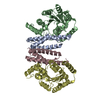
| ||||||||
| 2 | 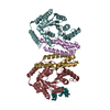
| ||||||||
| 3 | 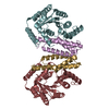
| ||||||||
| Unit cell |
|
-Components
| #1: Protein/peptide | Mass: 2826.475 Da / Num. of mol.: 1 Source method: isolated from a genetically manipulated source Source: (gene. exp.) Streptococcus pneumoniae (bacteria) / Plasmid: pET28b / Production host: Escherichia coli (E. coli) / Strain (production host): BL21(DE3)/Codon Plus-RIL | ||
|---|---|---|---|
| #2: Protein | Mass: 18267.666 Da / Num. of mol.: 4 Source method: isolated from a genetically manipulated source Source: (gene. exp.) Streptococcus pneumoniae (bacteria) / Strain: TIGR4 / Plasmid: pET28b / Production host: Escherichia coli (E. coli) / Strain (production host): BL21(DE3)/Codon Plus-RIL / References: UniProt: Q97QZ2#3: Protein | Mass: 29106.096 Da / Num. of mol.: 4 Source method: isolated from a genetically manipulated source Source: (gene. exp.) Streptococcus pneumoniae (bacteria) / Plasmid: pET28b / Production host: Escherichia coli (E. coli) / Strain (production host): BL21(DE3)/Codon Plus-RIL / References: UniProt: Q97QZ1 |
-Experimental details
-Experiment
| Experiment | Method: X-RAY DIFFRACTION / Number of used crystals: 1 |
|---|
- Sample preparation
| Crystal | Density Matthews: 2.74 Å3/Da / Density % sol: 55.09 % |
|---|---|
| Crystal grow | Temperature: 293 K / Method: vapor diffusion, hanging drop / pH: 6 Details: 12.5-15% (v/v) iso-propanol, 100 mM MES-NaOH, 6% (v/v) dioxane (30% (v/v)), pH 6.0, VAPOR DIFFUSION, HANGING DROP, temperature 293K |
-Data collection
| Diffraction | Mean temperature: 90 K |
|---|---|
| Diffraction source | Source: SYNCHROTRON / Site: SLS  / Beamline: X10SA / Wavelength: 1.007466 / Beamline: X10SA / Wavelength: 1.007466 |
| Detector | Type: MARMOSAIC 225 mm CCD / Detector: CCD / Date: Aug 17, 2006 |
| Radiation | Monochromator: Si(111) / Protocol: SINGLE WAVELENGTH / Monochromatic (M) / Laue (L): M / Scattering type: x-ray |
| Radiation wavelength | Wavelength: 1.007466 Å / Relative weight: 1 |
| Reflection | Resolution: 3.2→47.67 Å / Num. all: 34352 / Num. obs: 34352 / % possible obs: 95.3 % / Observed criterion σ(F): 3 / Redundancy: 5.7 % / Biso Wilson estimate: 76.2 Å2 / Rmerge(I) obs: 0.06 / Net I/σ(I): 20.8 |
| Reflection shell | Resolution: 3.2→3.3 Å / Rmerge(I) obs: 0.387 / Mean I/σ(I) obs: 3.1 / Num. unique all: 4708 / % possible all: 85.5 |
- Processing
| Software |
| ||||||||||||||||||||||||||||||||||||||||||||||||||||||||||||||||||||||||||||||||||||||||||||||||||||||||||||||||||||||||||||||||||||||||||||||||||||||
|---|---|---|---|---|---|---|---|---|---|---|---|---|---|---|---|---|---|---|---|---|---|---|---|---|---|---|---|---|---|---|---|---|---|---|---|---|---|---|---|---|---|---|---|---|---|---|---|---|---|---|---|---|---|---|---|---|---|---|---|---|---|---|---|---|---|---|---|---|---|---|---|---|---|---|---|---|---|---|---|---|---|---|---|---|---|---|---|---|---|---|---|---|---|---|---|---|---|---|---|---|---|---|---|---|---|---|---|---|---|---|---|---|---|---|---|---|---|---|---|---|---|---|---|---|---|---|---|---|---|---|---|---|---|---|---|---|---|---|---|---|---|---|---|---|---|---|---|---|---|---|---|
| Refinement | Method to determine structure: SAD / Resolution: 3.2→47.67 Å / Cor.coef. Fo:Fc: 0.932 / Cor.coef. Fo:Fc free: 0.893 / SU B: 55.632 / SU ML: 0.426 / TLS residual ADP flag: LIKELY RESIDUAL / Cross valid method: THROUGHOUT / ESU R Free: 0.52 / Stereochemistry target values: MAXIMUM LIKELIHOOD
| ||||||||||||||||||||||||||||||||||||||||||||||||||||||||||||||||||||||||||||||||||||||||||||||||||||||||||||||||||||||||||||||||||||||||||||||||||||||
| Solvent computation | Ion probe radii: 0.8 Å / Shrinkage radii: 0.8 Å / VDW probe radii: 1.2 Å / Solvent model: MASK | ||||||||||||||||||||||||||||||||||||||||||||||||||||||||||||||||||||||||||||||||||||||||||||||||||||||||||||||||||||||||||||||||||||||||||||||||||||||
| Displacement parameters | Biso mean: 91.268 Å2
| ||||||||||||||||||||||||||||||||||||||||||||||||||||||||||||||||||||||||||||||||||||||||||||||||||||||||||||||||||||||||||||||||||||||||||||||||||||||
| Refinement step | Cycle: LAST / Resolution: 3.2→47.67 Å
| ||||||||||||||||||||||||||||||||||||||||||||||||||||||||||||||||||||||||||||||||||||||||||||||||||||||||||||||||||||||||||||||||||||||||||||||||||||||
| Refine LS restraints |
| ||||||||||||||||||||||||||||||||||||||||||||||||||||||||||||||||||||||||||||||||||||||||||||||||||||||||||||||||||||||||||||||||||||||||||||||||||||||
| LS refinement shell | Resolution: 3.2→3.283 Å / Total num. of bins used: 20
| ||||||||||||||||||||||||||||||||||||||||||||||||||||||||||||||||||||||||||||||||||||||||||||||||||||||||||||||||||||||||||||||||||||||||||||||||||||||
| Refinement TLS params. | Method: refined / Refine-ID: X-RAY DIFFRACTION
| ||||||||||||||||||||||||||||||||||||||||||||||||||||||||||||||||||||||||||||||||||||||||||||||||||||||||||||||||||||||||||||||||||||||||||||||||||||||
| Refinement TLS group |
|
