 Movie
Movie Controller


+ Open data

- Basic information
| Entry | Database: PDB / ID: 2ouc | ||||||
|---|---|---|---|---|---|---|---|
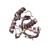
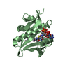
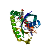
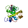
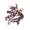
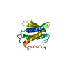
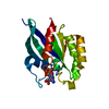
| Title | Crystal structure of the MAP kinase binding domain of MKP5 | ||||||
 Components Components | Dual specificity protein phosphatase 10 | ||||||
 Keywords Keywords |  HYDROLASE / Rhodanese fold HYDROLASE / Rhodanese fold | ||||||
| Function / homology |  Function and homology information Function and homology informationnegative regulation of epithelium regeneration / MAP kinase phosphatase activity / MAP kinase tyrosine phosphatase activity / protein tyrosine/threonine phosphatase activity / MAP kinase tyrosine/serine/threonine phosphatase activity / regulation of adaptive immune response / regulation of brown fat cell differentiation / negative regulation of p38MAPK cascade / negative regulation of epithelial cell migration / peptidyl-threonine dephosphorylation ...negative regulation of epithelium regeneration / MAP kinase phosphatase activity / MAP kinase tyrosine phosphatase activity / protein tyrosine/threonine phosphatase activity / MAP kinase tyrosine/serine/threonine phosphatase activity / regulation of adaptive immune response / regulation of brown fat cell differentiation / negative regulation of p38MAPK cascade / negative regulation of epithelial cell migration / peptidyl-threonine dephosphorylation / negative regulation of oligodendrocyte differentiation / Signaling by MAPK mutants / negative regulation of JUN kinase activity / RAF-independent MAPK1/3 activation / JUN kinase binding / negative regulation of JNK cascade / positive regulation of regulatory T cell differentiation / myosin phosphatase activity / mitogen-activated protein kinase p38 binding / peptidyl-tyrosine dephosphorylation involved in inactivation of protein kinase activity / protein-serine/threonine phosphatase / oligodendrocyte differentiation / phosphatase activity / negative regulation of respiratory burst involved in inflammatory response / stress-activated MAPK cascade / dephosphorylation / protein-tyrosine-phosphatase / negative regulation of cell migration / negative regulation of ERK1 and ERK2 cascade / Negative regulation of MAPK pathway / negative regulation of epithelial cell proliferation / response to lipopolysaccharide / nucleoplasm / nucleus / cytosol / cytoplasmSimilarity search - Function | ||||||
| Biological species |  Homo sapiens (human) Homo sapiens (human) | ||||||
| Method | X-RAY DIFFRACTION / SYNCHROTRON / SAD / Resolution: 2.2 Å | ||||||
 Authors Authors | Tao, X. / Tong, L. | ||||||
 Citation Citation | Journal: Protein Sci. / Year: 2007 Title: Crystal structure of the MAP kinase binding domain and the catalytic domain of human MKP5. Authors: Tao, X. / Tong, L. | ||||||
| History |
|
- Structure visualization
| Structure viewer | Molecule: MolmilJmol/JSmol |
|---|
- Downloads & links
-Download
| PDBx/mmCIF format | 2ouc.cif.gz | 65.3 KB | Display | PDBx/mmCIF format |
|---|---|---|---|---|
| PDB format | pdb2ouc.ent.gz | 49.3 KB | Display | PDB format |
| PDBx/mmJSON format | 2ouc.json.gz | Tree view | PDBx/mmJSON format | |
| Others |  Other downloads Other downloads |
-Validation report
| Arichive directory | https://data.pdbj.org/pub/pdb/validation_reports/ou/2oucftp://data.pdbj.org/pub/pdb/validation_reports/ou/2ouc | HTTPS FTP |
|---|
-Related structure data


-Links
 PDBj
PDBj


- Assembly
| Deposited unit | 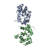
| ||||||||
|---|---|---|---|---|---|---|---|---|---|
| 1 | 
| ||||||||
| 2 | 
| ||||||||
| Unit cell |
|
-Components
| #1: Protein | Mass: 16233.819 Da / Num. of mol.: 2 / Fragment: Rhodanese domain (Residues 148-287) Source method: isolated from a genetically manipulated source Source: (gene. exp.) Homo sapiens (human) / Gene: MKP5 / Production host:  Escherichia coli (E. coli) Escherichia coli (E. coli)References: UniProt: Q9Y6W6, protein-tyrosine-phosphatase, protein-serine/threonine phosphatase#2: Water | ChemComp-HOH / | Water Mass: 18.015 Da / Num. of mol.: 115 / Source method: isolated from a natural source / Formula: H2O Mass: 18.015 Da / Num. of mol.: 115 / Source method: isolated from a natural source / Formula: H2O |
|---|
-Experimental details
-Experiment
| Experiment | Method: X-RAY DIFFRACTION / Number of used crystals: 1 |
|---|
- Sample preparation
| Crystal | Density Matthews: 2.46 Å3/Da / Density % sol: 50.09 % |
|---|---|
| Crystal grow | Temperature: 277 K / Method: vapor diffusion, sitting drop / pH: 4.5 Details: 100 mM sodium acetate (pH 4.5), and 2.3-2.5 M ammonium acetate, VAPOR DIFFUSION, SITTING DROP, temperature 277K |
-Data collection
| Diffraction | Mean temperature: 100 K |
|---|---|
| Diffraction source | Source: SYNCHROTRON / Site: NSLS  / Beamline: X4A / Wavelength: 0.9798 / Beamline: X4A / Wavelength: 0.9798 |
| Detector | Type: ADSC QUANTUM 4 / Detector: CCD / Details: MIRRORS |
| Radiation | Monochromator: SI(111) / Protocol: SINGLE WAVELENGTH / Monochromatic (M) / Laue (L): M / Scattering type: x-ray |
| Radiation wavelength | Wavelength: 0.9798 Å / Relative weight: 1 |
| Reflection | Resolution: 2.2→30 Å / Num. obs: 31415 / % possible obs: 97.7 % / Redundancy: 3.5 % / Biso Wilson estimate: 22.9 Å2 / Rmerge(I) obs: 0.05 / Net I/σ(I): 22.1234 |
| Reflection shell | Resolution: 2.2→2.28 Å / Redundancy: 2.8 % / Rmerge(I) obs: 0.263 / Mean I/σ(I) obs: 3.411 / % possible all: 88.4 |
- Processing
| Software |
| ||||||||||||||||||||||||||||||||||||||||||||||||||||||||||||||||||||||||||||||||
|---|---|---|---|---|---|---|---|---|---|---|---|---|---|---|---|---|---|---|---|---|---|---|---|---|---|---|---|---|---|---|---|---|---|---|---|---|---|---|---|---|---|---|---|---|---|---|---|---|---|---|---|---|---|---|---|---|---|---|---|---|---|---|---|---|---|---|---|---|---|---|---|---|---|---|---|---|---|---|---|---|---|
| Refinement | Method to determine structure: SAD / Resolution: 2.2→19.99 Å / Rfactor Rfree error: 0.006 / Data cutoff high absF: 579830.03 / Data cutoff low absF: 0 / Isotropic thermal model: RESTRAINED / Cross valid method: THROUGHOUT / σ(F): 1
| ||||||||||||||||||||||||||||||||||||||||||||||||||||||||||||||||||||||||||||||||
| Solvent computation | Solvent model: FLAT MODEL / Bsol: 54.5309 Å2 / ksol: 0.357953 e/Å3 | ||||||||||||||||||||||||||||||||||||||||||||||||||||||||||||||||||||||||||||||||
| Displacement parameters | Biso mean: 42.3 Å2
| ||||||||||||||||||||||||||||||||||||||||||||||||||||||||||||||||||||||||||||||||
| Refine analyze |
| ||||||||||||||||||||||||||||||||||||||||||||||||||||||||||||||||||||||||||||||||
| Refinement step | Cycle: LAST / Resolution: 2.2→19.99 Å
| ||||||||||||||||||||||||||||||||||||||||||||||||||||||||||||||||||||||||||||||||
| Refine LS restraints |
| ||||||||||||||||||||||||||||||||||||||||||||||||||||||||||||||||||||||||||||||||
| LS refinement shell | Resolution: 2.2→2.28 Å / Rfactor Rfree error: 0.02 / Total num. of bins used: 10
| ||||||||||||||||||||||||||||||||||||||||||||||||||||||||||||||||||||||||||||||||
| Xplor file |
|
