 Movie
Movie Controller


+ Open data

- Basic information
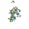
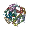
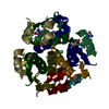
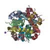
| Entry | Database: PDB / ID: 2omi | ||||||
|---|---|---|---|---|---|---|---|
| Title | Structure of human insulin cocrystallized with protamine | ||||||
 Components Components |
| ||||||
 Keywords Keywords |  HORMONE / insulin NPH microcrystals HORMONE / insulin NPH microcrystals | ||||||
| Function / homology |  Function and homology information Function and homology informationnegative regulation of NAD(P)H oxidase activity / negative regulation of glycogen catabolic process / regulation of cellular amino acid metabolic process / Signaling by Insulin receptor / IRS activation / nitric oxide-cGMP-mediated signaling / negative regulation of fatty acid metabolic process / Insulin processing / negative regulation of feeding behavior / regulation of protein secretion ...negative regulation of NAD(P)H oxidase activity / negative regulation of glycogen catabolic process / regulation of cellular amino acid metabolic process / Signaling by Insulin receptor / IRS activation / nitric oxide-cGMP-mediated signaling / negative regulation of fatty acid metabolic process / Insulin processing / negative regulation of feeding behavior / regulation of protein secretion / positive regulation of peptide hormone secretion / Regulation of gene expression in beta cells / positive regulation of respiratory burst / positive regulation of dendritic spine maintenance / alpha-beta T cell activation / negative regulation of acute inflammatory response / negative regulation of respiratory burst involved in inflammatory response / negative regulation of protein secretion / fatty acid homeostasis / Synthesis, secretion, and deacylation of Ghrelin / positive regulation of glycogen biosynthetic process / positive regulation of lipid biosynthetic process / Signal attenuation / FOXO-mediated transcription of oxidative stress, metabolic and neuronal genes / negative regulation of gluconeogenesis / positive regulation of nitric oxide mediated signal transduction / regulation of protein localization to plasma membrane / COPI-mediated anterograde transport / negative regulation of lipid catabolic process / negative regulation of oxidative stress-induced intrinsic apoptotic signaling pathway / negative regulation of reactive oxygen species biosynthetic process / positive regulation of insulin receptor signaling pathway / transport vesicle / positive regulation of protein autophosphorylation / Insulin receptor recycling / insulin-like growth factor receptor binding / NPAS4 regulates expression of target genes / positive regulation of protein metabolic process / neuron projection maintenance / endoplasmic reticulum-Golgi intermediate compartment membrane / positive regulation of brown fat cell differentiation / activation of protein kinase B activity / positive regulation of glycolytic process / Insulin receptor signalling cascade / positive regulation of mitotic nuclear division / Regulation of insulin secretion / positive regulation of nitric-oxide synthase activity / positive regulation of long-term synaptic potentiation / endosome lumen / positive regulation of cytokine production / acute-phase response / positive regulation of protein secretion / regulation of transmembrane transporter activity / positive regulation of cell differentiation / positive regulation of glucose import / negative regulation of proteolysis / regulation of synaptic plasticity / wound healing / insulin receptor binding / negative regulation of protein catabolic process / positive regulation of neuron projection development / hormone activity / cognition / Golgi lumen / vasodilation / positive regulation of protein localization to nucleus / glucose metabolic process / regulation of protein localization / glucose homeostasis / cell-cell signaling / insulin receptor signaling pathway / positive regulation of NF-kappaB transcription factor activity / PI5P, PP2A and IER3 Regulate PI3K/AKT Signaling / positive regulation of cell growth / secretory granule lumen / protease binding / positive regulation of MAPK cascade / positive regulation of phosphatidylinositol 3-kinase/protein kinase B signal transduction / positive regulation of cell migration / G protein-coupled receptor signaling pathway / Amyloid fiber formation / endoplasmic reticulum lumen / Golgi membrane / negative regulation of gene expression / positive regulation of cell population proliferation / positive regulation of gene expression / regulation of DNA-templated transcription / extracellular space / extracellular region / identical protein bindingSimilarity search - Function | ||||||
| Biological species |  Homo sapiens (human) Homo sapiens (human) | ||||||
| Method | X-RAY DIFFRACTION / SYNCHROTRON / MOLECULAR REPLACEMENT / Resolution: 2.24 Å | ||||||
 Authors Authors | Norrman, M. / Schluckebier, G. | ||||||
 Citation Citation | Journal: EUR.J.PHARM.SCI. / Year: 2007 Title: Structural characterization of insulin NPH formulations. Authors: Norrman, M. / Hubalek, F. / Schluckebier, G. | ||||||
| History |
|
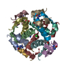
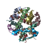
- Structure visualization
| Structure viewer | Molecule: MolmilJmol/JSmol |
|---|
- Downloads & links
-Download
| PDBx/mmCIF format | 2omi.cif.gz | 73.3 KB | Display | PDBx/mmCIF format |
|---|---|---|---|---|
| PDB format | pdb2omi.ent.gz | 56.6 KB | Display | PDB format |
| PDBx/mmJSON format | 2omi.json.gz | Tree view | PDBx/mmJSON format | |
| Others |  Other downloads Other downloads |
-Validation report
| Arichive directory | https://data.pdbj.org/pub/pdb/validation_reports/om/2omiftp://data.pdbj.org/pub/pdb/validation_reports/om/2omi | HTTPS FTP |
|---|
-Related structure data


-Links
 PDBj
PDBj


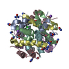
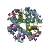
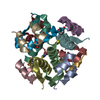
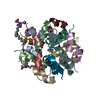
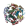
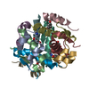
- Assembly
| Deposited unit | 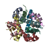
| ||||||||
|---|---|---|---|---|---|---|---|---|---|
| 1 |
| ||||||||
| Unit cell |
|
-Components
-Protein/peptide , 2 types, 12 molecules ACEGIKBDFHJL
| #1: Protein/peptide | Mass: 2383.698 Da / Num. of mol.: 6 / Source method: isolated from a natural source / Source: (natural) Homo sapiens (human) / References: UniProt: P01308#2: Protein/peptide | Mass: 3433.953 Da / Num. of mol.: 6 / Source method: isolated from a natural source / Source: (natural) Homo sapiens (human) / References: UniProt: P01308 |
|---|
-Non-polymers , 4 types, 120 molecules 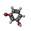


| #3: Chemical | ChemComp-RCO / Resorcinol Mass: 110.111 Da / Num. of mol.: 6 / Source method: obtained synthetically / Formula: C6H6O2 Mass: 110.111 Da / Num. of mol.: 6 / Source method: obtained synthetically / Formula: C6H6O2#4: Chemical |  Mass: 65.409 Da / Num. of mol.: 2 / Source method: obtained synthetically / Formula: Zn Mass: 65.409 Da / Num. of mol.: 2 / Source method: obtained synthetically / Formula: Zn#5: Chemical | Chloride Mass: 35.453 Da / Num. of mol.: 2 / Source method: obtained synthetically / Formula: Cl Mass: 35.453 Da / Num. of mol.: 2 / Source method: obtained synthetically / Formula: Cl#6: Water | ChemComp-HOH / | WaterMass: 18.015 Da / Num. of mol.: 110 / Source method: isolated from a natural source / Formula: H2O |
|---|
-Experimental details
-Experiment
| Experiment | Method: X-RAY DIFFRACTION / Number of used crystals: 1 |
|---|
- Sample preparation
| Crystal | Density Matthews: 2.3 Å3/Da / Density % sol: 46.63 % |
|---|---|
| Crystal grow | Temperature: 291 K / Method: vapor diffusion, hanging drop / pH: 7.3 Details: 50mM resorcinol, 400mM NaCl, 1.0mg/ml protamine 30mM phosphate buffer, pH 7.3, VAPOR DIFFUSION, HANGING DROP, temperature 291K |
-Data collection
| Diffraction | Mean temperature: 100 K |
|---|---|
| Diffraction source | Source: SYNCHROTRON / Site: SLS  / Beamline: X06SA / Wavelength: 0.918 Å / Beamline: X06SA / Wavelength: 0.918 Å |
| Detector | Type: MAR CCD 165 mm / Detector: CCD / Date: Jun 23, 2005 |
| Radiation | Monochromator: LN2 cooled fixed-exit Si(111) monochromator / Protocol: SINGLE WAVELENGTH / Monochromatic (M) / Laue (L): M / Scattering type: x-ray |
| Radiation wavelength | Wavelength: 0.918 Å / Relative weight: 1 |
| Reflection | Resolution: 2.24→30 Å / Num. all: 16106 / Num. obs: 15881 / % possible obs: 98.6 % / Redundancy: 6.5 % / Biso Wilson estimate: 33.7 Å2 / Rmerge(I) obs: 0.107 / Net I/σ(I): 12.1 |
| Reflection shell | Resolution: 2.24→2.4 Å / Redundancy: 2.8 % / Rmerge(I) obs: 0.286 / Mean I/σ(I) obs: 4.1 / Num. unique all: 2781 / % possible all: 97.6 |
- Processing
| Software |
| ||||||||||||||||||||||||||||||||||||||||||||||||||||||||||||||||||||||||||||||||||||||||||||||||||||||||||||||||||||||||||||||||||||||||||||||||||||||||||||||||||||||||||
|---|---|---|---|---|---|---|---|---|---|---|---|---|---|---|---|---|---|---|---|---|---|---|---|---|---|---|---|---|---|---|---|---|---|---|---|---|---|---|---|---|---|---|---|---|---|---|---|---|---|---|---|---|---|---|---|---|---|---|---|---|---|---|---|---|---|---|---|---|---|---|---|---|---|---|---|---|---|---|---|---|---|---|---|---|---|---|---|---|---|---|---|---|---|---|---|---|---|---|---|---|---|---|---|---|---|---|---|---|---|---|---|---|---|---|---|---|---|---|---|---|---|---|---|---|---|---|---|---|---|---|---|---|---|---|---|---|---|---|---|---|---|---|---|---|---|---|---|---|---|---|---|---|---|---|---|---|---|---|---|---|---|---|---|---|---|---|---|---|---|---|---|
| Refinement | Method to determine structure: MOLECULAR REPLACEMENT Starting model: insulin hexamer R-conformation Resolution: 2.24→29.1 Å / Cor.coef. Fo:Fc: 0.93 / Cor.coef. Fo:Fc free: 0.878 / SU B: 6.045 / SU ML: 0.158 / Isotropic thermal model: isotropic / Cross valid method: THROUGHOUT / ESU R: 0.324 / ESU R Free: 0.251 / Stereochemistry target values: MAXIMUM LIKELIHOOD / Details: HYDROGENS HAVE BEEN ADDED IN THE RIDING POSITIONS
| ||||||||||||||||||||||||||||||||||||||||||||||||||||||||||||||||||||||||||||||||||||||||||||||||||||||||||||||||||||||||||||||||||||||||||||||||||||||||||||||||||||||||||
| Solvent computation | Ion probe radii: 0.8 Å / Shrinkage radii: 0.8 Å / VDW probe radii: 1.4 Å / Solvent model: MASK | ||||||||||||||||||||||||||||||||||||||||||||||||||||||||||||||||||||||||||||||||||||||||||||||||||||||||||||||||||||||||||||||||||||||||||||||||||||||||||||||||||||||||||
| Displacement parameters | Biso mean: 26.301 Å2
| ||||||||||||||||||||||||||||||||||||||||||||||||||||||||||||||||||||||||||||||||||||||||||||||||||||||||||||||||||||||||||||||||||||||||||||||||||||||||||||||||||||||||||
| Refinement step | Cycle: LAST / Resolution: 2.24→29.1 Å
| ||||||||||||||||||||||||||||||||||||||||||||||||||||||||||||||||||||||||||||||||||||||||||||||||||||||||||||||||||||||||||||||||||||||||||||||||||||||||||||||||||||||||||
| Refine LS restraints |
| ||||||||||||||||||||||||||||||||||||||||||||||||||||||||||||||||||||||||||||||||||||||||||||||||||||||||||||||||||||||||||||||||||||||||||||||||||||||||||||||||||||||||||
| LS refinement shell | Resolution: 2.24→2.296 Å / Total num. of bins used: 20
|
