 Movie
Movie Controller

[English] 日本語
 Yorodumi
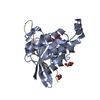
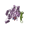
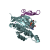
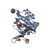
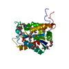
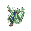
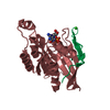
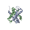
- PDB-2odb: The crystal structure of human cdc42 in complex with the CRIB dom... -
+ Open data

- Basic information
| Entry | Database: PDB / ID: 2odb | ||||||
|---|---|---|---|---|---|---|---|
| Title | The crystal structure of human cdc42 in complex with the CRIB domain of human p21-activated kinase 6 (PAK6) | ||||||
 Components Components |
| ||||||
 Keywords Keywords |  PROTEIN BINDING / small GTPase / CRIB / kinase / protein-protein complex / Structural Genomics / Structural Genomics Consortium / SGC PROTEIN BINDING / small GTPase / CRIB / kinase / protein-protein complex / Structural Genomics / Structural Genomics Consortium / SGC | ||||||
| Function / homology |  Function and homology information Function and homology informationGBD domain binding / submandibular salivary gland formation / actin filament branching / Golgi transport complex / positive regulation of pinocytosis / modification of synaptic structure / endothelin receptor signaling pathway involved in heart process / Cdc42 protein signal transduction / cardiac neural crest cell migration involved in outflow tract morphogenesis / positive regulation of synapse structural plasticity ...GBD domain binding / submandibular salivary gland formation / actin filament branching / Golgi transport complex / positive regulation of pinocytosis / modification of synaptic structure / endothelin receptor signaling pathway involved in heart process / Cdc42 protein signal transduction / cardiac neural crest cell migration involved in outflow tract morphogenesis / positive regulation of synapse structural plasticity / dendritic cell migration / storage vacuole / apolipoprotein A-I receptor binding / positive regulation of epithelial cell proliferation involved in lung morphogenesis / neuron fate determination / modulation by host of viral process / organelle transport along microtubule / regulation of attachment of spindle microtubules to kinetochore / positive regulation of pseudopodium assembly / Inactivation of CDC42 and RAC1 / cardiac conduction system development / neuron projection arborization / GTP-dependent protein binding / regulation of filopodium assembly / establishment of Golgi localization / leading edge membrane / neuropilin signaling pathway / positive regulation of intracellular protein transport / cell junction assembly / filopodium assembly / establishment of epithelial cell apical/basal polarity / regulation of modification of postsynaptic structure / dendritic spine morphogenesis / mitogen-activated protein kinase kinase kinase binding / embryonic heart tube development / thioesterase binding / regulation of stress fiber assembly / RHOD GTPase cycle / RHO GTPases activate KTN1 / regulation of lamellipodium assembly / Activation of RAC1 / nuclear migration / DCC mediated attractive signaling / adherens junction organization / sprouting angiogenesis / Wnt signaling pathway, planar cell polarity pathway / CD28 dependent Vav1 pathway / regulation of postsynapse organization / positive regulation of filopodium assembly / neuron projection extension / regulation of mitotic nuclear division / establishment or maintenance of cell polarity / phagocytosis, engulfment / RHOV GTPase cycle / heart contraction / Myogenesis / RHOJ GTPase cycle / Golgi organization / RHOQ GTPase cycle / positive regulation of cytokinesis / RHO GTPases activate PAKs / regulation of MAPK cascade / RHOH GTPase cycle / CDC42 GTPase cycle / RHOU GTPase cycle / macrophage differentiation / RHOG GTPase cycle / RHO GTPases Activate WASPs and WAVEs / RAC3 GTPase cycle / RAC2 GTPase cycle / RHO GTPases activate IQGAPs / spindle midzone / positive regulation of DNA replication / negative regulation of protein-containing complex assembly / phagocytic vesicle / positive regulation of lamellipodium assembly / positive regulation of substrate adhesion-dependent cell spreading / cytoskeleton organization / positive regulation of stress fiber assembly / GPVI-mediated activation cascade / RAC1 GTPase cycle / EPHB-mediated forward signaling / substantia nigra development / Gene and protein expression by JAK-STAT signaling after Interleukin-12 stimulation / small monomeric GTPase / G protein activity / locomotory behavior / filopodium / learning / secretory granule / actin filament organization / integrin-mediated signaling pathway / RHO GTPases Activate Formins / regulation of actin cytoskeleton organization / FCGR3A-mediated phagocytosis / EGFR downregulation / positive regulation of JNK cascade / MAPK6/MAPK4 signaling / Schaffer collateral - CA1 synapse / protein localizationSimilarity search - Function | ||||||
| Biological species |  Homo sapiens (human) Homo sapiens (human) | ||||||
| Method | X-RAY DIFFRACTION / SYNCHROTRON / MOLECULAR REPLACEMENT / Resolution: 2.4 Å | ||||||
 Authors Authors | Ugochukwu, E. / Yang, X. / Elkins, J. / Soundararajan, M. / Pike, A.C.W. / Eswaran, J. / Burgess, N. / Debreczeni, J.E. / Sundstrom, M. / Arrowsmith, C. ...Ugochukwu, E. / Yang, X. / Elkins, J. / Soundararajan, M. / Pike, A.C.W. / Eswaran, J. / Burgess, N. / Debreczeni, J.E. / Sundstrom, M. / Arrowsmith, C. / Weigelt, J. / Edwards, A. / Gileadi, O. / von Delft, F. / Knapp, S. / Doyle, D. / Structural Genomics Consortium (SGC) | ||||||
 Citation Citation | Journal: To be Published Title: The crystal structure of human cdc42 in complex with the CRIB domain of human p21-activated kinase 6 (PAK6) Authors: Yang, X. / Ugochukwu, E. / Elkins, J. / Soundararajan, M. / Eswaran, J. / Pike, A.C.W. / Burgess, N. / Debreczeni, J.E. / Gileadi, O. / Knapp, S. / Doyle, D. | ||||||
| History |
| ||||||
| Remark 999 | sequence The sequence of human CDC42 isoform 1 is not available in uniprot database at the time of processing. |
- Structure visualization
| Structure viewer | Molecule: MolmilJmol/JSmol |
|---|
- Downloads & links
-Download
| PDBx/mmCIF format | 2odb.cif.gz | 60.1 KB | Display | PDBx/mmCIF format |
|---|---|---|---|---|
| PDB format | pdb2odb.ent.gz | 41.7 KB | Display | PDB format |
| PDBx/mmJSON format | 2odb.json.gz | Tree view | PDBx/mmJSON format | |
| Others |  Other downloads Other downloads |
-Validation report
| Arichive directory | https://data.pdbj.org/pub/pdb/validation_reports/od/2odbftp://data.pdbj.org/pub/pdb/validation_reports/od/2odb | HTTPS FTP |
|---|
-Related structure data
| Related structure data | 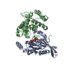 1grnS S: Starting model for refinement |
|---|---|
| Similar structure data |


-Links
 PDBj
PDBj


- Assembly
| Deposited unit | 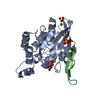
| ||||||||
|---|---|---|---|---|---|---|---|---|---|
| 1 |
| ||||||||
| 2 | 
| ||||||||
| 3 | 
| ||||||||
| 4 | 
| ||||||||
| Unit cell |
| ||||||||
| Components on special symmetry positions |
| ||||||||
| Details | One monomer of each protein in the asymmetric unit |
-Components
-Protein / Protein/peptide , 2 types, 2 molecules AB
| #1: Protein | Mass: 21370.660 Da / Num. of mol.: 1 Source method: isolated from a genetically manipulated source Source: (gene. exp.) Homo sapiens (human) / Gene: CDC42 / Plasmid: pNIC-Bsa4 / Species (production host): Escherichia coli / Production host:  Escherichia coli BL21(DE3) (bacteria) / Strain (production host): BL21 (DE3) / References: UniProt: P60953 Escherichia coli BL21(DE3) (bacteria) / Strain (production host): BL21 (DE3) / References: UniProt: P60953 |
|---|---|
| #2: Protein/peptide | Mass: 4066.510 Da / Num. of mol.: 1 / Fragment: PAK6 CRIB domain / Source method: obtained synthetically / References: UniProt: Q9NQU5 |
-Non-polymers , 5 types, 50 molecules 


| #3: Chemical | ChemComp-CL / Chloride Mass: 35.453 Da / Num. of mol.: 1 / Source method: obtained synthetically / Formula: Cl Mass: 35.453 Da / Num. of mol.: 1 / Source method: obtained synthetically / Formula: Cl | ||||
|---|---|---|---|---|---|
| #4: Chemical | ChemComp-MG /  Mass: 24.305 Da / Num. of mol.: 1 / Source method: obtained synthetically / Formula: Mg Mass: 24.305 Da / Num. of mol.: 1 / Source method: obtained synthetically / Formula: Mg | ||||
| #5: Chemical | Sulfate Mass: 96.063 Da / Num. of mol.: 3 / Source method: obtained synthetically / Formula: SO4 Mass: 96.063 Da / Num. of mol.: 3 / Source method: obtained synthetically / Formula: SO4#6: Chemical | ChemComp-GCP / |  Mass: 521.208 Da / Num. of mol.: 1 / Source method: obtained synthetically / Formula: C11H18N5O13P3 / Comment: GMP-PCP, energy-carrying molecule analogue*YM Mass: 521.208 Da / Num. of mol.: 1 / Source method: obtained synthetically / Formula: C11H18N5O13P3 / Comment: GMP-PCP, energy-carrying molecule analogue*YM#7: Water | ChemComp-HOH / | WaterMass: 18.015 Da / Num. of mol.: 44 / Source method: isolated from a natural source / Formula: H2O |
-Experimental details
-Experiment
| Experiment | Method: X-RAY DIFFRACTION / Number of used crystals: 1 |
|---|
- Sample preparation
| Crystal | Density Matthews: 3.44 Å3/Da / Density % sol: 64.2 % |
|---|---|
| Crystal grow | Temperature: 293 K / Method: vapor diffusion, sitting drop / pH: 4.5 Details: 0.1M acetate, 2M (NH4)2SO4, pH 4.5, VAPOR DIFFUSION, SITTING DROP, temperature 293K |
-Data collection
| Diffraction | Mean temperature: 100 K |
|---|---|
| Diffraction source | Source: SYNCHROTRON / Site: SLS  / Beamline: X10SA / Wavelength: 0.97956 / Beamline: X10SA / Wavelength: 0.97956 |
| Detector | Type: MARMOSAIC 225 mm CCD / Detector: CCD / Date: Nov 26, 2006 |
| Radiation | Protocol: SINGLE WAVELENGTH / Monochromatic (M) / Laue (L): M / Scattering type: x-ray |
| Radiation wavelength | Wavelength: 0.97956 Å / Relative weight: 1 |
| Reflection | Resolution: 2.4→40.59 Å / Num. obs: 14484 / % possible obs: 100 % / Observed criterion σ(F): 0 / Observed criterion σ(I): 0 / Rmerge(I) obs: 0.071 / Rsym value: 0.071 |
| Reflection shell | Resolution: 2.4→2.53 Å / Rmerge(I) obs: 0.502 / Rsym value: 0.502 / % possible all: 100 |
- Processing
| Software |
| ||||||||||||||||||||||||||||||||||||||||||||||||||||||||||||||||||||||||||||||||||||||||||||||||||||||||||||||||||||||||||||||||||||||||||||||||||||||||||||||||||||||||||
|---|---|---|---|---|---|---|---|---|---|---|---|---|---|---|---|---|---|---|---|---|---|---|---|---|---|---|---|---|---|---|---|---|---|---|---|---|---|---|---|---|---|---|---|---|---|---|---|---|---|---|---|---|---|---|---|---|---|---|---|---|---|---|---|---|---|---|---|---|---|---|---|---|---|---|---|---|---|---|---|---|---|---|---|---|---|---|---|---|---|---|---|---|---|---|---|---|---|---|---|---|---|---|---|---|---|---|---|---|---|---|---|---|---|---|---|---|---|---|---|---|---|---|---|---|---|---|---|---|---|---|---|---|---|---|---|---|---|---|---|---|---|---|---|---|---|---|---|---|---|---|---|---|---|---|---|---|---|---|---|---|---|---|---|---|---|---|---|---|---|---|---|
| Refinement | Method to determine structure: MOLECULAR REPLACEMENT Starting model: pdb entry 1GRN Resolution: 2.4→40.59 Å / Cor.coef. Fo:Fc: 0.953 / Cor.coef. Fo:Fc free: 0.946 / SU B: 14.923 / SU ML: 0.182 / TLS residual ADP flag: LIKELY RESIDUAL / Cross valid method: THROUGHOUT / σ(F): 0 / ESU R: 0.249 / ESU R Free: 0.2 / Stereochemistry target values: MAXIMUM LIKELIHOOD / Details: HYDROGENS HAVE BEEN ADDED IN THE RIDING POSITIONS
| ||||||||||||||||||||||||||||||||||||||||||||||||||||||||||||||||||||||||||||||||||||||||||||||||||||||||||||||||||||||||||||||||||||||||||||||||||||||||||||||||||||||||||
| Solvent computation | Ion probe radii: 0.8 Å / Shrinkage radii: 0.8 Å / VDW probe radii: 1.4 Å / Solvent model: MASK | ||||||||||||||||||||||||||||||||||||||||||||||||||||||||||||||||||||||||||||||||||||||||||||||||||||||||||||||||||||||||||||||||||||||||||||||||||||||||||||||||||||||||||
| Displacement parameters | Biso mean: 47.158 Å2
| ||||||||||||||||||||||||||||||||||||||||||||||||||||||||||||||||||||||||||||||||||||||||||||||||||||||||||||||||||||||||||||||||||||||||||||||||||||||||||||||||||||||||||
| Refinement step | Cycle: LAST / Resolution: 2.4→40.59 Å
| ||||||||||||||||||||||||||||||||||||||||||||||||||||||||||||||||||||||||||||||||||||||||||||||||||||||||||||||||||||||||||||||||||||||||||||||||||||||||||||||||||||||||||
| Refine LS restraints |
| ||||||||||||||||||||||||||||||||||||||||||||||||||||||||||||||||||||||||||||||||||||||||||||||||||||||||||||||||||||||||||||||||||||||||||||||||||||||||||||||||||||||||||
| LS refinement shell | Resolution: 2.4→2.462 Å / Total num. of bins used: 20
| ||||||||||||||||||||||||||||||||||||||||||||||||||||||||||||||||||||||||||||||||||||||||||||||||||||||||||||||||||||||||||||||||||||||||||||||||||||||||||||||||||||||||||
| Refinement TLS params. | Method: refined / Refine-ID: X-RAY DIFFRACTION
| ||||||||||||||||||||||||||||||||||||||||||||||||||||||||||||||||||||||||||||||||||||||||||||||||||||||||||||||||||||||||||||||||||||||||||||||||||||||||||||||||||||||||||
| Refinement TLS group |
|
