 Movie
Movie Controller

[English] 日本語
 Yorodumi
- PDB-2o6l: Crystal Structure of the UDP-Glucuronic Acid Binding Domain of th... -
+ Open data

- Basic information
| Entry | Database: PDB / ID: 2o6l | ||||||
|---|---|---|---|---|---|---|---|
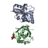
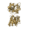
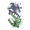
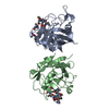
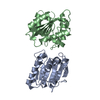
| Title | Crystal Structure of the UDP-Glucuronic Acid Binding Domain of the Human Drug Metabolizing UDP-Glucuronosyltransferase 2B7 | ||||||
 Components Components | UDP-glucuronosyltransferase 2B7 | ||||||
 Keywords Keywords |  TRANSFERASE / Drug metabolism / rossman / MAD / enzyme / nucleotide binding / sugar / UDP-glucuronosyltransferase / UGT TRANSFERASE / Drug metabolism / rossman / MAD / enzyme / nucleotide binding / sugar / UDP-glucuronosyltransferase / UGT | ||||||
| Function / homology |  Function and homology informationglucuronosyltransferase / cellular glucuronidation / Glucuronidation / glucuronosyltransferase activity / androgen metabolic process / lipid metabolic process / membrane => GO:0016020 / intracellular membrane-bounded organelle / endoplasmic reticulum membrane / membrane Function and homology informationglucuronosyltransferase / cellular glucuronidation / Glucuronidation / glucuronosyltransferase activity / androgen metabolic process / lipid metabolic process / membrane => GO:0016020 / intracellular membrane-bounded organelle / endoplasmic reticulum membrane / membraneSimilarity search - Function | ||||||
| Biological species |  Homo sapiens (human) Homo sapiens (human) | ||||||
| Method | X-RAY DIFFRACTION / SYNCHROTRON / MAD / Resolution: 1.8 Å | ||||||
 Authors Authors | Miley, M.J. / Redinbo, M.R. | ||||||
 Citation Citation | Journal: J.Mol.Biol. / Year: 2007 Title: Crystal Structure of the Cofactor-Binding Domain of the Human Phase II Drug-Metabolism Enzyme UDP-Glucuronosyltransferase 2B7. Authors: Miley, M.J. / Zielinska, A.K. / Keenan, J.E. / Bratton, S.M. / Radominska-Pandya, A. / Redinbo, M.R. | ||||||
| History |
|
- Structure visualization
| Structure viewer | Molecule: MolmilJmol/JSmol |
|---|
- Downloads & links
-Download
| PDBx/mmCIF format | 2o6l.cif.gz | 84.5 KB | Display | PDBx/mmCIF format |
|---|---|---|---|---|
| PDB format | pdb2o6l.ent.gz | 65.3 KB | Display | PDB format |
| PDBx/mmJSON format | 2o6l.json.gz | Tree view | PDBx/mmJSON format | |
| Others |  Other downloads Other downloads |
-Validation report
| Arichive directory | https://data.pdbj.org/pub/pdb/validation_reports/o6/2o6lftp://data.pdbj.org/pub/pdb/validation_reports/o6/2o6l | HTTPS FTP |
|---|
-Related structure data
| Similar structure data |
|---|


-Links
 PDBj
PDBj- Assembly
| Deposited unit | 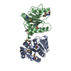
| ||||||||||||||||||
|---|---|---|---|---|---|---|---|---|---|---|---|---|---|---|---|---|---|---|---|
| 1 |
| ||||||||||||||||||
| Unit cell |
| ||||||||||||||||||
| Components on special symmetry positions |
|
-Components
| #1: Protein | Mass: 19125.721 Da / Num. of mol.: 2 Fragment: UDP-Glucuronic acid binding domain, residues 285-472 Source method: isolated from a genetically manipulated source Source: (gene. exp.) Homo sapiens (human) / Gene: UGT2B7 / Production host:  Escherichia coli (E. coli) / Strain (production host): B834(DE3) / References: UniProt: P16662, glucuronosyltransferase Escherichia coli (E. coli) / Strain (production host): B834(DE3) / References: UniProt: P16662, glucuronosyltransferase#2: Water | ChemComp-HOH / | Water Mass: 18.015 Da / Num. of mol.: 283 / Source method: isolated from a natural source / Formula: H2O Mass: 18.015 Da / Num. of mol.: 283 / Source method: isolated from a natural source / Formula: H2O |
|---|
-Experimental details
-Experiment
| Experiment | Method: X-RAY DIFFRACTION / Number of used crystals: 1 |
|---|
- Sample preparation
| Crystal | Density Matthews: 2.05 Å3/Da / Density % sol: 39.97 % |
|---|---|
| Crystal grow | Temperature: 295 K / Method: vapor diffusion, hanging drop / pH: 7.4 Details: 12-16% PEG 4000, 100 mM K2CO3, 100 mM Tris, pH 7.4, VAPOR DIFFUSION, HANGING DROP, temperature 295K |
-Data collection
| Diffraction |
| |||||||||||||||||||||||||
|---|---|---|---|---|---|---|---|---|---|---|---|---|---|---|---|---|---|---|---|---|---|---|---|---|---|---|
| Diffraction source |
| |||||||||||||||||||||||||
| Detector | Type: MARMOSAIC 300 mm CCD / Detector: CCD / Date: Feb 12, 2006 | |||||||||||||||||||||||||
| Radiation | Monochromator: SAGITALLY FOCUSED Si(220) / Protocol: MAD / Monochromatic (M) / Laue (L): M / Scattering type: x-ray | |||||||||||||||||||||||||
| Radiation wavelength |
| |||||||||||||||||||||||||
| Reflection | Resolution: 1.8→50 Å / Num. all: 34624 / Num. obs: 33813 / % possible obs: 97.7 % / Observed criterion σ(F): 0 / Observed criterion σ(I): 0 / Redundancy: 14.4 % / Biso Wilson estimate: 21.2 Å2 / Rsym value: 5.6 / Net I/σ(I): 50 | |||||||||||||||||||||||||
| Reflection shell | Resolution: 1.8→1.86 Å / Redundancy: 13.4 % / Mean I/σ(I) obs: 6.5 / Num. unique all: 3171 / Rsym value: 39.2 / % possible all: 94.3 |

- Processing
| Software |
| ||||||||||||||||||||||||||||||||||||
|---|---|---|---|---|---|---|---|---|---|---|---|---|---|---|---|---|---|---|---|---|---|---|---|---|---|---|---|---|---|---|---|---|---|---|---|---|---|
| Refinement | Method to determine structure: MAD / Resolution: 1.8→48.52 Å / Rfactor Rfree error: 0.004 / Data cutoff high absF: 103739.1 / Data cutoff low absF: 0 / Isotropic thermal model: RESTRAINED / Cross valid method: THROUGHOUT / σ(F): 0 / σ(I): 0 / Stereochemistry target values: CNS Details: MAD 3 wavelengths were used to solve structure to 2.1 angstroms (multi-wavelength method, wavelengths 0.97925, 0.97942, 0.97174). Once model traced and refined, 1.80 angstrom dataset was ...Details: MAD 3 wavelengths were used to solve structure to 2.1 angstroms (multi-wavelength method, wavelengths 0.97925, 0.97942, 0.97174). Once model traced and refined, 1.80 angstrom dataset was used for final refinements (low-remote-dataset, wavelength 0.98089).
| ||||||||||||||||||||||||||||||||||||
| Solvent computation | Solvent model: FLAT MODEL / Bsol: 44.1013 Å2 / ksol: 0.325832 e/Å3 | ||||||||||||||||||||||||||||||||||||
| Displacement parameters | Biso mean: 29.9 Å2
| ||||||||||||||||||||||||||||||||||||
| Refine analyze |
| ||||||||||||||||||||||||||||||||||||
| Refinement step | Cycle: LAST / Resolution: 1.8→48.52 Å
| ||||||||||||||||||||||||||||||||||||
| Refine LS restraints |
| ||||||||||||||||||||||||||||||||||||
| LS refinement shell | Resolution: 1.8→1.91 Å / Rfactor Rfree error: 0.016 / Total num. of bins used: 6
| ||||||||||||||||||||||||||||||||||||
| Xplor file |
|
