 Movie
Movie Controller

[English] 日本語
 Yorodumi
- PDB-2nyf: Crystal structure of phenylalanine ammonia-lyase from Nostoc punc... -
+ Open data

- Basic information
| Entry | Database: PDB / ID: 2nyf | |||||||||
|---|---|---|---|---|---|---|---|---|---|---|
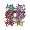
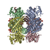
| Title | Crystal structure of phenylalanine ammonia-lyase from Nostoc punctiforme | |||||||||
 Components Components | Nostoc punctiforme phenylalanine ammonia lyase | |||||||||
 Keywords Keywords |  LYASE / Methylidene imidazolone prosthetic group (autocatalytically formed by internal tripeptide segment Ala167-Ser168-Gly169) LYASE / Methylidene imidazolone prosthetic group (autocatalytically formed by internal tripeptide segment Ala167-Ser168-Gly169) | |||||||||
| Function / homology |  Function and homology informationphenylalanine ammonia-lyase / cinnamic acid biosynthetic process / phenylalanine ammonia-lyase activity / phenylpropanoid biosynthetic process / aromatic amino acid metabolic process / protein homotetramerization / cytoplasm Function and homology informationphenylalanine ammonia-lyase / cinnamic acid biosynthetic process / phenylalanine ammonia-lyase activity / phenylpropanoid biosynthetic process / aromatic amino acid metabolic process / protein homotetramerization / cytoplasmSimilarity search - Function | |||||||||
| Biological species |  Nostoc punctiforme (bacteria) Nostoc punctiforme (bacteria) | |||||||||
| Method | X-RAY DIFFRACTION / SYNCHROTRON / MOLECULAR REPLACEMENT / Resolution: 2.5 Å | |||||||||
 Authors Authors | Louie, G.V. / Moffitt, M.C. / Bowman, M.E. / Pence, J. / Noel, J.P. / Moore, B.S. | |||||||||
 Citation Citation | Journal: Biochemistry / Year: 2007 Title: Discovery of Two Cyanobacterial Phenylalanine Ammonia Lyases: Kinetic and Structural Characterization. Authors: Moffitt, M.C. / Louie, G.V. / Bowman, M.E. / Pence, J. / Noel, J.P. / Moore, B.S. | |||||||||
| History |
| |||||||||
| Remark 600 | HETEROGEN Residue MDO is autocatalytically formed by internal tripeptide segment Ala167-Ser168-Gly169 | |||||||||
| Remark 999 | Sequence No suitable database references were found at time of processing |
- Structure visualization
| Structure viewer | Molecule: MolmilJmol/JSmol |
|---|
- Downloads & links
-Download
| PDBx/mmCIF format | 2nyf.cif.gz | 110.9 KB | Display | PDBx/mmCIF format |
|---|---|---|---|---|
| PDB format | pdb2nyf.ent.gz | 85 KB | Display | PDB format |
| PDBx/mmJSON format | 2nyf.json.gz | Tree view | PDBx/mmJSON format | |
| Others |  Other downloads Other downloads |
-Validation report
| Arichive directory | https://data.pdbj.org/pub/pdb/validation_reports/ny/2nyfftp://data.pdbj.org/pub/pdb/validation_reports/ny/2nyf | HTTPS FTP |
|---|
-Related structure data


-Links
 PDBj
PDBj

- Assembly
| Deposited unit | 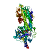
| |||||||||
|---|---|---|---|---|---|---|---|---|---|---|
| 1 | 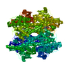
| |||||||||
| Unit cell |
| |||||||||
| Components on special symmetry positions |
| |||||||||
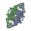
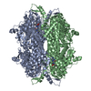
| Details | The 222 symmetric homotetramer is generated by crystallographic symmetry. |
-Components
| #1: Protein | Mass: 62264.680 Da / Num. of mol.: 1 Source method: isolated from a genetically manipulated source Source: (gene. exp.) Nostoc punctiforme (bacteria) / Strain: PCC 73102 / Plasmid: pHIS8 / Species (production host): Escherichia coli / Production host: Escherichia coli BL21 (bacteria) / Strain (production host): BL21 / References: UniProt: B2J528*PLUS, histidine ammonia-lyase |
|---|---|
| #2: Water | ChemComp-HOH / Water Mass: 18.015 Da / Num. of mol.: 31 / Source method: isolated from a natural source / Formula: H2O Mass: 18.015 Da / Num. of mol.: 31 / Source method: isolated from a natural source / Formula: H2O |
-Experimental details
-Experiment
| Experiment | Method: X-RAY DIFFRACTION / Number of used crystals: 1 |
|---|
- Sample preparation
| Crystal | Density Matthews: 2.06 Å3/Da / Density % sol: 40.31 % |
|---|---|
| Crystal grow | Temperature: 277 K / Method: vapor diffusion, hanging drop / pH: 7 Details: 0.1 M MOPSO (pH 7.0), 21% (w/v) polyethylene glycol 8000, 0.2 M lithium sulfate, 2 mM dithiothreitol, VAPOR DIFFUSION, HANGING DROP, temperature 277K |
-Data collection
| Diffraction | Mean temperature: 100 K |
|---|---|
| Diffraction source | Source: SYNCHROTRON / Site: ALS  / Beamline: 8.2.2 / Wavelength: 1 Å / Beamline: 8.2.2 / Wavelength: 1 Å |
| Detector | Type: ADSC QUANTUM 315 / Detector: CCD / Date: Apr 1, 2006 |
| Radiation | Protocol: SINGLE WAVELENGTH / Monochromatic (M) / Laue (L): M / Scattering type: x-ray |
| Radiation wavelength | Wavelength: 1 Å / Relative weight: 1 |
| Reflection | Resolution: 2.5→100 Å / Num. all: 18191 / Num. obs: 16902 / % possible obs: 92.9 % / Redundancy: 4.8 % / Biso Wilson estimate: 33.3 Å2 / Rmerge(I) obs: 0.098 / Net I/σ(I): 12.56 |
| Reflection shell | Resolution: 2.5→2.59 Å / Redundancy: 2.8 % / Rmerge(I) obs: 0.475 / Mean I/σ(I) obs: 2.8 / Num. unique all: 1809 / % possible all: 69.7 |
- Processing
| Software |
| |||||||||||||||||||||
|---|---|---|---|---|---|---|---|---|---|---|---|---|---|---|---|---|---|---|---|---|---|---|
| Refinement | Method to determine structure: MOLECULAR REPLACEMENT / Resolution: 2.5→100 Å / Isotropic thermal model: Isotropic / Cross valid method: Random / σ(F): 0 / σ(I): 0 / Stereochemistry target values: Engh & Huber
| |||||||||||||||||||||
| Displacement parameters | Biso mean: 47.1 Å2 | |||||||||||||||||||||
| Refinement step | Cycle: LAST / Resolution: 2.5→100 Å
| |||||||||||||||||||||
| Refine LS restraints |
| |||||||||||||||||||||
| LS refinement shell | Resolution: 2.5→2.61 Å /
|
