 Movie
Movie Controller

[English] 日本語
 Yorodumi
- PDB-2nvu: Structure of APPBP1-UBA3~NEDD8-NEDD8-MgATP-Ubc12(C111A), a trappe... -
+ Open data

- Basic information
| Entry | Database: PDB / ID: 2nvu | ||||||
|---|---|---|---|---|---|---|---|
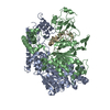
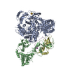
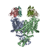
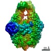
| Title | Structure of APPBP1-UBA3~NEDD8-NEDD8-MgATP-Ubc12(C111A), a trapped ubiquitin-like protein activation complex | ||||||
 Components Components |
| ||||||
 Keywords Keywords |  PROTEIN TURNOVER / LIGASE / Multifunction macromolecular complex / Ubiquitin / NEDD8 / E1 / E2 / ATP / Conformational change / thioester / switch / adenylation PROTEIN TURNOVER / LIGASE / Multifunction macromolecular complex / Ubiquitin / NEDD8 / E1 / E2 / ATP / Conformational change / thioester / switch / adenylation | ||||||
| Function / homology |  Function and homology information Function and homology informationE2 NEDD8-conjugating enzyme / NEDD8 conjugating enzyme activity / E1 NEDD8-activating enzyme / NEDD8 activating enzyme activity / endomitotic cell cycle / ubiquitin activating enzyme activity / NEDD8 transferase activity / regulation of proteolysis / mitotic DNA replication checkpoint signaling / protein neddylation ...E2 NEDD8-conjugating enzyme / NEDD8 conjugating enzyme activity / E1 NEDD8-activating enzyme / NEDD8 activating enzyme activity / endomitotic cell cycle / ubiquitin activating enzyme activity / NEDD8 transferase activity / regulation of proteolysis / mitotic DNA replication checkpoint signaling / protein neddylation / TGF-beta receptor signaling activates SMADs / anatomical structure morphogenesis / regulation of neuron apoptotic process / post-translational protein modification / NIK-->noncanonical NF-kB signaling / Dectin-1 mediated noncanonical NF-kB signaling / Iron uptake and transport / protein modification process / modification-dependent protein catabolic process / protein localization / protein tag activity / ubiquitin-protein transferase activity / UCH proteinases / positive regulation of neuron apoptotic process / Antigen processing: Ubiquitination & Proteasome degradation / Cargo recognition for clathrin-mediated endocytosis / Neddylation / ubiquitin-dependent protein catabolic process / neuron apoptotic process / regulation of apoptotic process / protein ubiquitination / regulation of cell cycle / protein heterodimerization activity / DNA damage response / ubiquitin protein ligase binding / regulation of transcription by RNA polymerase II / signal transduction / protein-containing complex / proteolysis / extracellular exosome / nucleoplasm / ATP binding / identical protein binding / nucleus / plasma membrane / cytosol / cytoplasmSimilarity search - Function | ||||||
| Biological species |  Homo sapiens (human) Homo sapiens (human) | ||||||
| Method | X-RAY DIFFRACTION / SYNCHROTRON / MOLECULAR REPLACEMENT / Resolution: 2.8 Å | ||||||
 Authors Authors | Huang, D.T. / Hunt, H.W. / Zhuang, M. / Ohi, M.D. / Holton, J.M. / Schulman, B.A. | ||||||
 Citation Citation | Journal: Nature / Year: 2007 Title: Basis for a ubiquitin-like protein thioester switch toggling E1-E2 affinity. Authors: Huang, D.T. / Hunt, H.W. / Zhuang, M. / Ohi, M.D. / Holton, J.M. / Schulman, B.A. | ||||||
| History |
| ||||||
| Remark 999 | SEQUENCE ENTITY 2 (CHAIN B) CONTAINS TWO DIFFERENT PROTEINS: MALTOSE BINDING PROTEIN (MBP, RESIDUES ...SEQUENCE ENTITY 2 (CHAIN B) CONTAINS TWO DIFFERENT PROTEINS: MALTOSE BINDING PROTEIN (MBP, RESIDUES 1001-1368) AND NEDD8-ACTIVATING ENZYME E1 CATALYTIC SUBUNIT (UBA3, RESIDUES 2012-2442) CONNECTED BY THE ALA-ALA-ALA LINKER (RESIDUES 1369-1371). THERE IS NO DATABASE SEQUENCE AVAILABLE AT UNIPROT FOR MBP AT THE TIME OF DEPOSITION. AUTHORS STATE THAT THE MBP SEQUENCE IS AVAILABLE ONLY IN NCBI DATABASE WITH ACCESSION NUMBERS AAB86559 OR 1R6Z_P. PROTEIN UBA3 HAS A DATABASE SEQUENCE REFERENCE IN UNIPROT WHICH IS STATED IN DBREF. ENTITY 2 ALSO CONTAINS N-TERMINAL CLONING ARTIFACT (RESIDUES MET-LYS-LEU, RESIDUES 998-1000) AND THREE MUTATIONS (E1360A,K1363A,D1364A). THE C-TERMINAL CARBON ATOM OF CHAIN "J" (RESIDUE 76) FORMS A THIOESTER LINK WITH CHAIN "B" BY THE SIDE CHAIN ATOM SG OF RESIDUE 2216 OF THE UBA3 MOIETY |
- Structure visualization
| Structure viewer | Molecule: MolmilJmol/JSmol |
|---|
- Downloads & links
-Download
| PDBx/mmCIF format | 2nvu.cif.gz | 339.5 KB | Display | PDBx/mmCIF format |
|---|---|---|---|---|
| PDB format | pdb2nvu.ent.gz | 266.6 KB | Display | PDB format |
| PDBx/mmJSON format | 2nvu.json.gz | Tree view | PDBx/mmJSON format | |
| Others |  Other downloads Other downloads |
-Validation report
| Arichive directory | https://data.pdbj.org/pub/pdb/validation_reports/nv/2nvuftp://data.pdbj.org/pub/pdb/validation_reports/nv/2nvu | HTTPS FTP |
|---|
-Related structure data


-Links
 PDBj
PDBj


- Assembly
| Deposited unit | 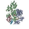
| ||||||||
|---|---|---|---|---|---|---|---|---|---|
| 1 |
| ||||||||
| Unit cell |
|
-Components
-Protein , 4 types, 5 molecules ABCIJ
| #1: Protein | Mass: 60459.227 Da / Num. of mol.: 1 Source method: isolated from a genetically manipulated source Source: (gene. exp.) Homo sapiens (human) / Gene: APPBP1 / Plasmid: pGEX4T-1 / Production host:  Escherichia coli (E. coli) / Strain (production host): BL21(DE3)GOLD / References: UniProt: Q13564 Escherichia coli (E. coli) / Strain (production host): BL21(DE3)GOLD / References: UniProt: Q13564 |
|---|---|
| #2: Protein | Mass: 89332.766 Da / Num. of mol.: 1 / Fragment: Residues 33-463 Source method: isolated from a genetically manipulated source Details: Fusion protein. Maltose binding protein (residues 1001-1371), and the NEDD8-activating enzyme E1 catalytic subunit (residues 2012-2442) is from a human source Source: (gene. exp.) Homo sapiens (human) / Gene: UBE1C, UBA3 / Plasmid: pGEX4T-1 / Production host: Escherichia coli (E. coli) / Strain (production host): BL21(DE3)GOLDReferences: UniProt: Q8TBC4, Ligases; Forming carbon-nitrogen bonds; Acid-amino-acid ligases (peptide synthases) |
| #3: Protein | Mass: 20665.666 Da / Num. of mol.: 1 / Mutation: del(S16-T20), C111A Source method: isolated from a genetically manipulated source Source: (gene. exp.) Homo sapiens (human) / Gene: UBE2M, UBC12 / Plasmid: pGEX4T-1 / Production host: Escherichia coli (E. coli) / Strain (production host): BL21(DE3)GOLDReferences: UniProt: P61081, Ligases; Forming carbon-nitrogen bonds; Acid-amino-acid ligases (peptide synthases) |
| #4: Protein | / Ubiquitin-like protein Nedd8 / Neddylin Mass: 8919.289 Da / Num. of mol.: 2 Source method: isolated from a genetically manipulated source Source: (gene. exp.) Homo sapiens (human) / Gene: NEDD8 / Plasmid: pGEX4T-1 / Production host: Escherichia coli (E. coli) / Strain (production host): BL21(DE3)GOLD / References: UniProt: Q15843 |
-Non-polymers , 4 types, 48 molecules 


| #5: Chemical | ChemComp-MG /  Mass: 24.305 Da / Num. of mol.: 1 / Source method: obtained synthetically / Formula: Mg Mass: 24.305 Da / Num. of mol.: 1 / Source method: obtained synthetically / Formula: Mg |
|---|---|
| #6: Chemical | ChemComp-ZN /  Mass: 65.409 Da / Num. of mol.: 1 / Source method: obtained synthetically / Formula: Zn Mass: 65.409 Da / Num. of mol.: 1 / Source method: obtained synthetically / Formula: Zn |
| #7: Chemical | ChemComp-ATP / Adenosine triphosphate Mass: 507.181 Da / Num. of mol.: 1 / Source method: obtained synthetically / Formula: C10H16N5O13P3 / Comment: ATP, energy-carrying molecule*YM Mass: 507.181 Da / Num. of mol.: 1 / Source method: obtained synthetically / Formula: C10H16N5O13P3 / Comment: ATP, energy-carrying molecule*YM |
| #8: Water | ChemComp-HOH / WaterMass: 18.015 Da / Num. of mol.: 45 / Source method: isolated from a natural source / Formula: H2O |
-Experimental details
-Experiment
| Experiment | Method: X-RAY DIFFRACTION / Number of used crystals: 1 |
|---|
- Sample preparation
| Crystal | Density Matthews: 3.57 Å3/Da / Density % sol: 65.59 % Description: AUTHORS SCREENED >900 CRYSTALS AND USED ALL THREE BEAMLINES TO OBTAIN NECESSARY INFORMATION FOR FINAL STRUCTURE DETERMINATION FROM SINGLE CRYSTAL AT APS BEAMLINE 22-ID |
|---|---|
| Crystal grow | Temperature: 291 K / pH: 7 Details: 17% v/v PEG 3350, 0.1 M HEPES pH 7.0, 0.2 M Disodium tartrate, VAPOR DIFFUSION, HANGING DROP, temperature 291K, pH 7.00 |
-Data collection
| Diffraction | Mean temperature: 100 K |
|---|---|
| Diffraction source | Source: SYNCHROTRON / Site: APS  / Beamline: 22-ID / Wavelength: 0.9793 / Beamline: 22-ID / Wavelength: 0.9793 |
| Detector | Type: MARMOSAIC 300 mm CCD / Detector: CCD / Date: Jun 16, 2006 |
| Radiation | Protocol: SINGLE WAVELENGTH / Monochromatic (M) / Laue (L): M / Scattering type: x-ray |
| Radiation wavelength | Wavelength: 0.9793 Å / Relative weight: 1 |
| Reflection | Resolution: 2.8→50 Å / Num. obs: 65807 / % possible obs: 95.7 % / Observed criterion σ(I): 0 / Redundancy: 7.6 % / Rsym value: 0.134 / Net I/σ(I): 25.7 |
- Processing
| Software |
| ||||||||||||||||||||||||||||||||||||||||||||||||||||||||||||
|---|---|---|---|---|---|---|---|---|---|---|---|---|---|---|---|---|---|---|---|---|---|---|---|---|---|---|---|---|---|---|---|---|---|---|---|---|---|---|---|---|---|---|---|---|---|---|---|---|---|---|---|---|---|---|---|---|---|---|---|---|---|
| Refinement | Method to determine structure: MOLECULAR REPLACEMENT Starting model: PDB ENTRIES 1R4M, 1Y8X Resolution: 2.8→50 Å / Isotropic thermal model: ISOTROPIC / Cross valid method: FREE R / σ(F): 0 / Stereochemistry target values: ENGH & HUBER
| ||||||||||||||||||||||||||||||||||||||||||||||||||||||||||||
| Solvent computation | Solvent model: FLAT MODEL / Bsol: 32.29 Å2 | ||||||||||||||||||||||||||||||||||||||||||||||||||||||||||||
| Displacement parameters | Biso mean: 83.78 Å2
| ||||||||||||||||||||||||||||||||||||||||||||||||||||||||||||
| Refinement step | Cycle: LAST / Resolution: 2.8→50 Å
| ||||||||||||||||||||||||||||||||||||||||||||||||||||||||||||
| Refine LS restraints |
| ||||||||||||||||||||||||||||||||||||||||||||||||||||||||||||
| Xplor file |
|
