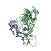
| 1 | Sample 1: 0.2 mM [U-15N; U-13C; U-2H] PARP-1 1, 0.2 mM DNA (45-MER), 50 mM [U-2H] TRIS, 1 mM [U-2H] DTT, 0.1 mM ZnSO4, 95% H2O/5% D2O | 95% H2O/5% D2O| 2 | Sample 2: 0.2 mM [U-15N; U-13C; U-70% 2H] PARP-1 1, 0.2 mM DNA (45-MER), 50 mM [U-2H] TRIS, 1 mM [U-2H] DTT, 0.1 mM ZnSO4, 95% H2O/5% D2O | 95% H2O/5% D2O| 3 | Sample 3: 0.2 mM [U-98% 2H; U-98% 15N] PARP-1 1, 0.2 mM DNA (45-MER), 50 mM [U-2H] TRIS, 1 mM [U-2H] DTT, 0.1 mM ZnSO4, 95% H2O/5% D2O | 95% H2O/5% D2O| 4 | Sample 4a: 0.2 MM PARP-1 1-214, Uniform [2H,15N,13C], back-labeled with [1H,13C] in the delta-methyl groups of Ile and all methyl groups of Leu and Val residues, using sodium [4-13C, 3,3-2H2] alpha-ketobutyrate and sodium [3- 2H, 4,4'-13C2] alpha-ketoisovalerate as precursors to maximize protonation of methyl groups, for use in NOE experiments; sodium [3-2H, 4,4'-13C2] alpha-ketoisovalerate was prepared from sodium [4,4'-13C2] alpha-ketoisovalerate by exchange with 2H2O at pH 12.5 and 45 C for 3 hrs. 0.2 mM DNA (45-MER), 50 mM [U-2H] TRIS, 1 mM [U-2H] DTT, 0.1 mM ZnSO4, 95% H2O/5% D2O | 95% H2O/5% D2O| 5 | Sample 4b: 0.2 MM PARP-1 1-214, Uniform [2H,15N,13C], back-labeled with [1H,13C] in the delta-methyl groups of Ile and all methyl groups of Leu and Val residues, using sodium [3,3-2H2,13C4] alpha-ketobutyrate and sodium [3- 2H,13C5] alpha-ketoisovalerate as precursors to produce linear chains of 13C in the sidechains of Val and Leu, for use in assignment experiments to link methyl signals to C-alpha signals. 0.2 mM DNA (45-MER), 50 mM [U-2H] TRIS, 1 mM [U-2H] DTT, 0.1 mM ZnSO4, 100% D2O | 100% D2O| 6 | Sample 5: 0.2 MM PARP-1 1-214, Uniform [2H,15N,13C], back-labeled with [1H,13C] in the methyl groups of Met residues in addition to Ile, Leu and Val methyl groups as in sample 4a. 0.2 mM DNA (45-MER), 50 mM [U-2H] TRIS, 1 mM [U-2H] DTT, 0.1 mM ZnSO4, 100% D2O | 100% D2O| 7 | Sample 6: 0.2 MM PARP-1 1-214, Uniform [2H,15N,13C]; back-labeled with [1H,13C] in the methyl groups of Ile, Leu and Val methyl groups as in sample 4a and [1H,13C,15N] Phe residues. 0.2 mM DNA (45-MER), 50 mM [U-2H] TRIS, 1 mM [U-2H] DTT, 0.1 mM ZnSO4, 100% D2O | 100% D2O| 8 | Sample 7: 0.2 MM PARP-1 1-214, Sortase ligated, block-labelled sample. (N.B. residues 103 and 104 of WT sequence deleted, additional residues LPETGGG inserted between residues 102 and 105; this sample was not used for making any assignments of residues in this region, which is in the flexible linker between domains). Labelling for residues 1-102 (and LPET of insertion): uniform [1H,12C,15N]. Labelling for residues 105-214 (and GGG of insertion): [2H,15N,13C] back-labeled with [1H,13C] Ile, Leu Val and Met methyl groups labeled as in sample 5. 0.2 mM DNA (45-MER), 50 mM [U-2H] TRIS, 1 mM [U-2H] DTT, 0.1 mM ZnSO4, 100% D2O | 100% D2O| 9 | Sample 8: 0.2 MM PARP-1 1-214, Sortase ligated, block-labelled sample. (N.B. residues 103 and 104 of WT sequence deleted, additional residues LPETGGG inserted between residues 102 and 105; this sample was not used for making any assignments of residues in this region, which is in the flexible linker between domains). Labelling for residues 1-102 (and LPET of insertion): uniform [1H,12C,15N]. Labelling for residues 105-214 (and GGG of insertion): [2H,15N,13C] back labeled with [1H,13C] Met methyl groups as in sample 5 and [13C,15N,1H] Arg residues. 0.2 mM DNA (45-MER), 50 mM [U-2H] TRIS, 1 mM [U-2H] DTT, 0.1 mM ZnSO4, 100% D2O | 100% D2O| 10 | Sample 9: 0.2 MM PARP-1 1-214, Sortase ligated, block-labelled sample. (N.B. residues 103 and 104 of WT sequence deleted, additional residues LPETGGG inserted between residues 102 and 105; this sample was not used for making any assignments of residues in this region, which is in the flexible linker between domains). Labelling for residues 1-102 (and LPET of insertion): [2H,15N,13C] back-labeled with Ile, Leu and Val methyl groups labeled as in sample 4a and [1H,13C,15N] Arg residues. Labelling for residues 105-214 (and GGG of insertion): uniform [1H,12C,15N]. 0.2 mM DNA (45-MER), 50 mM [U-2H] TRIS, 1 mM [U-2H] DTT, 0.1 mM ZnSO4, 95% H2O/5% D2O | 95% H2O/5% D2O| 11 | Sample 10: 0.2 MM PARP-1 1-214, Sortase ligated, block-labelled sample. (N.B. residues 103 and 104 of WT sequence deleted, additional residues LPETGGG inserted between residues 102 and 105; this sample was not used for making any assignments of residues in this region, which is in the flexible linker between domains). Labelling for residues 1-102 (and LPET of insertion): [2H,15N,13C] back-labeled with Ile, Leu and Val methyl groups labeled as in sample 4a and [1H,13C,15N] Phe residues. Labelling for residues 105-214 (and GGG of insertion): uniform [1H,12C,15N]. 0.2 mM DNA (45-MER), 50 mM [U-2H] TRIS, 1 mM [U-2H] DTT, 0.1 mM ZnSO4, 100% D2O | 100% D2O| 12 | Sample 11: 0.2 mM see Sample details section PARP-1 1, 0.2 mM DNA (45-MER), 50 mM [U-2H] TRIS, 1 mM [U-2H] DTT, 0.1 mM ZnSO4, 100% D2O | 100% D2O| 13 | Sample 12: 0.2 mM DNA (45-MER), 50 mM [U-2H] TRIS, 1 mM [U-2H] DTT, 0.1 mM ZnSO4, 100% D2O | 100% D2O| 14 | Sample 13: 0.2 mM DNA (45-MER), 50 mM [U-2H] TRIS, 1 mM [U-2H] DTT, 0.1 mM ZnSO4, 95% H2O/5% D2O | 95% H2O/5% D2O| 15 | Sample 14: 0.2 mM [U-15N; U-13C; U-2H] PARP-1 1, 0.2 mM DNA (45-MER), 50 mM [U-2H] TRIS, 1 mM [U-2H] DTT, 0.1 mM ZnSO4, 100% D2O | 100% D2O| 16 | Sample 15: 0.2 mM [U-15N; U-13C; U-2H] PARP-1 1, 50 mM [U-2H] TRIS, 1 mM [U-2H] DTT, 0.1 uM ZnSO4, 200 mM sodium chloride, 95% H2O/5% D2O | 95% H2O/5% D2O| 17 | Sample 16: 0.2 mM [U-15N; U-13C; U-2H] PARP-1 1, 0.2 mM DNA (45-MER), 50 mM [U-2H] TRIS, 1 mM [U-2H] DTT, 0.1 uM ZnSO4, 200 mM sodium chloride, 95% H2O/5% D2O | 95% H2O/5% D2O | | | | | | | | | | | | | | | | |
 Yorodumi
 Open data
 Basic information
 Components
 Keywords
 TRANSFERASE
 Function and homology information
 regulation of base-excision repair / positive regulation of myofibroblast differentiation / NAD+-protein-tyrosine ADP-ribosyltransferase activity / NAD+-protein-histidine ADP-ribosyltransferase activity / negative regulation of ATP biosynthetic process / carbohydrate biosynthetic process / regulation of circadian sleep/wake cycle, non-REM sleep ...NAD+-histone H2BS6 serine ADP-ribosyltransferase activity / NAD+-histone H3S10 serine ADP-ribosyltransferase activity / NAD+-histone H2BE35 glutamate ADP-ribosyltransferase activity /
  regulation of base-excision repair / positive regulation of myofibroblast differentiation / NAD+-protein-tyrosine ADP-ribosyltransferase activity / NAD+-protein-histidine ADP-ribosyltransferase activity / negative regulation of ATP biosynthetic process / carbohydrate biosynthetic process / regulation of circadian sleep/wake cycle, non-REM sleep / positive regulation of single strand break repair / vRNA Synthesis / NAD+-protein-serine ADP-ribosyltransferase activity / negative regulation of adipose tissue development / NAD DNA ADP-ribosyltransferase activity / NAD+- protein-aspartate ADP-ribosyltransferase activity / NAD+-protein-glutamate ADP-ribosyltransferase activity / DNA ADP-ribosylation / mitochondrial DNA metabolic process / regulation of oxidative stress-induced neuron intrinsic apoptotic signaling pathway / signal transduction involved in regulation of gene expression / replication fork reversal / positive regulation of necroptotic process /
  ATP generation from poly-ADP-D-ribose /
  regulation of catalytic activity / HDR through MMEJ (alt-NHEJ) / transcription regulator activator activity / : / positive regulation of DNA-templated transcription, elongation /
  NAD+ ADP-ribosyltransferase / cellular response to zinc ion / negative regulation of telomere maintenance via telomere lengthening / positive regulation of intracellular estrogen receptor signaling pathway / protein auto-ADP-ribosylation / positive regulation of mitochondrial depolarization / response to aldosterone / mitochondrial DNA repair / negative regulation of cGAS/STING signaling pathway / positive regulation of double-strand break repair via homologous recombination / protein poly-ADP-ribosylation / positive regulation of cardiac muscle hypertrophy / nuclear replication fork / negative regulation of transcription elongation by RNA polymerase II / site of DNA damage / NAD+-protein ADP-ribosyltransferase activity /
  R-SMAD binding / positive regulation of SMAD protein signal transduction / macrophage differentiation / protein autoprocessing /
  decidualization /
  NAD+ ADP-ribosyltransferase activity /
  Transferases; Glycosyltransferases; Pentosyltransferases /
  nucleosome binding / POLB-Dependent Long Patch Base Excision Repair / SUMOylation of DNA damage response and repair proteins / protein localization to chromatin /
  telomere maintenance / negative regulation of innate immune response / mitochondrion organization /
  nucleotidyltransferase activity / cellular response to nerve growth factor stimulus / transforming growth factor beta receptor signaling pathway / nuclear estrogen receptor binding /
  protein-DNA complex / response to gamma radiation / Downregulation of SMAD2/3:SMAD4 transcriptional activity / DNA Damage Recognition in GG-NER / protein modification process / Dual Incision in GG-NER / Formation of Incision Complex in GG-NER / positive regulation of protein localization to nucleus /
  histone deacetylase binding / cellular response to insulin stimulus / cellular response to amyloid-beta / cellular response to UV / NAD binding /
  regulation of protein localization / double-strand break repair / site of double-strand break /
  nuclear envelope / cellular response to oxidative stress / positive regulation of canonical NF-kappaB signal transduction /
  transcription regulator complex / RNA polymerase II-specific DNA-binding transcription factor binding / transcription by RNA polymerase II / damaged DNA binding /
  chromosome, telomeric region /
  nuclear body /
  DNA repair /
  innate immune response / negative regulation of DNA-templated transcription / apoptotic process / DNA damage response /
  chromatin binding /
  ubiquitin protein ligase binding /
  chromatin /
  nucleolus /
  protein kinase binding / negative regulation of transcription by RNA polymerase II /
  enzyme binding
 Homo sapiens (human)
 SOLUTION NMR /
  simulated annealing
  Authors
 Citation
 Journal: Mol.Cell / Year: 2015
 Structure visualization
 Molmil
 Jmol/JSmol
 Downloads & links
 Download
 2n8a.cif.gz
 PDBx/mmCIF format
 pdb2n8a.ent.gz
 PDB format
 2n8a.json.gz
 PDBx/mmJSON format
 Other downloads
 https://data.pdbj.org/pub/pdb/validation_reports/n8/2n8a
 ftp://data.pdbj.org/pub/pdb/validation_reports/n8/2n8a
 Links
Links Assembly
 Components
 Homo sapiens (human) / Gene: PARP1, ADPRT, PPOL / Production host:
 Escherichia coli (E. coli) / Strain (production host): BL21 / Variant (production host): DE3 / References: UniProt: P09874,
  NAD+ ADP-ribosyltransferase
 SOLUTION NMR
 Sample preparation
 Movie
Movie Controller





 PDBj
PDBj


