 Movie
Movie Controller

[English] 日本語
 Yorodumi
- PDB-2ltq: High resolution structure of DsbB C41S by joint calculation with ... -
+ Open data

- Basic information
| Entry | Database: PDB / ID: 2ltq | |||||||||
|---|---|---|---|---|---|---|---|---|---|---|
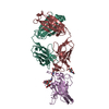
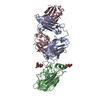
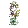
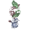
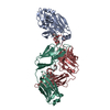
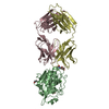
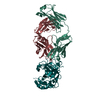
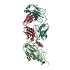
| Title | High resolution structure of DsbB C41S by joint calculation with solid-state NMR and X-ray data | |||||||||
 Components Components |
| |||||||||
 Keywords Keywords |  MEMBRANE PROTEIN / OXIDOREDUCTASE / DISULFIDE BOND / REDOX-ACTIVE CENTER / CELL INNER MEMBRANE / CELL MEMBRANE / CHAPERONE / ELECTRON TRANSPORT / MEMBRANE / TRANSMEMBRANE / TRANSPORT MEMBRANE PROTEIN / OXIDOREDUCTASE / DISULFIDE BOND / REDOX-ACTIVE CENTER / CELL INNER MEMBRANE / CELL MEMBRANE / CHAPERONE / ELECTRON TRANSPORT / MEMBRANE / TRANSMEMBRANE / TRANSPORT | |||||||||
| Function / homology |  Function and homology information Function and homology informationoxidoreductase activity, acting on a sulfur group of donors, quinone or similar compound as acceptor / ubiquinone binding / protein-disulfide reductase activity / protein folding / response to heat / electron transfer activity / plasma membraneSimilarity search - Function | |||||||||
| Biological species |  Escherichia coli (E. coli) Escherichia coli (E. coli) Mus musculus (house mouse) Mus musculus (house mouse) | |||||||||
| Method | SOLID-STATE NMR / simulated annealing | |||||||||
| Model details | lowest energy, model 1 | |||||||||
 Authors Authors | Tang, M. / Sperling, L.J. / Schwieters, C.D. / Nesbitt, A.E. / Gennis, R.B. / Rienstra, C.M. | |||||||||
 Citation Citation | Journal: J.Mol.Biol. / Year: 2013 Title: Structure of the Disulfide Bond Generating Membrane Protein DsbB in the Lipid Bilayer. Authors: Tang, M. / Nesbitt, A.E. / Sperling, L.J. / Berthold, D.A. / Schwieters, C.D. / Gennis, R.B. / Rienstra, C.M. | |||||||||
| History |
|
- Structure visualization
| Structure viewer | Molecule: MolmilJmol/JSmol |
|---|
- Downloads & links
-Download
| PDBx/mmCIF format | 2ltq.cif.gz | 3.4 MB | Display | PDBx/mmCIF format |
|---|---|---|---|---|
| PDB format | pdb2ltq.ent.gz | 2.9 MB | Display | PDB format |
| PDBx/mmJSON format | 2ltq.json.gz | Tree view | PDBx/mmJSON format | |
| Others |  Other downloads Other downloads |
-Validation report
| Arichive directory | https://data.pdbj.org/pub/pdb/validation_reports/lt/2ltqftp://data.pdbj.org/pub/pdb/validation_reports/lt/2ltq | HTTPS FTP |
|---|
-Related structure data
| Related structure data | |
|---|---|
| Similar structure data | |
| Other databases |


-Links
 PDBj
PDBj


- Assembly
| Deposited unit | 
| |||||||||
|---|---|---|---|---|---|---|---|---|---|---|
| 1 | 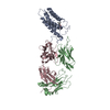
| |||||||||
| 2 | 
| |||||||||
| NMR ensembles |
|
-Components
| #1: Protein | / Disulfide oxidoreductase Mass: 20106.982 Da / Num. of mol.: 2 / Mutation: C8A,C41S,C49V Source method: isolated from a genetically manipulated source Source: (gene. exp.) Escherichia coli (strain K12) (bacteria)Strain: K12 / Gene: dsbB, roxB, ycgA, b1185, JW5182 / Production host: Escherichia coli (E. coli) / References: UniProt: P0A6M2#2: Antibody | Fragment antigen-bindingMass: 26364.410 Da / Num. of mol.: 2 / Source method: isolated from a natural source Details: FAB FRAGMENT IS OBTAINED BY PAPAIN DIGESTION OF A SELECTED MONOCLONAL ANTIBODY Source: (natural) Mus musculus (house mouse)#3: Antibody | Fragment antigen-bindingMass: 23666.453 Da / Num. of mol.: 2 / Source method: isolated from a natural source Details: FAB FRAGMENT IS OBTAINED BY PAPAIN DIGESTION OF A SELECTED MONOCLONAL ANTIBODY Source: (natural) Mus musculus (house mouse)#4: Chemical |   Mass: 727.109 Da / Num. of mol.: 2 / Source method: obtained synthetically / Formula: C49H74O4 Mass: 727.109 Da / Num. of mol.: 2 / Source method: obtained synthetically / Formula: C49H74O4 |
|---|
-Experimental details
-Experiment
| Experiment | Method: SOLID-STATE NMR | ||||||||||||||||||||||||
|---|---|---|---|---|---|---|---|---|---|---|---|---|---|---|---|---|---|---|---|---|---|---|---|---|---|
| NMR experiment |
| ||||||||||||||||||||||||
| NMR details | Text: Chemical shifts assignments and CC correlations provide dihedral angle and distance restraints. |
- Sample preparation
| Details |
| ||||||||||||||||||||||||||||||||||||||||
|---|---|---|---|---|---|---|---|---|---|---|---|---|---|---|---|---|---|---|---|---|---|---|---|---|---|---|---|---|---|---|---|---|---|---|---|---|---|---|---|---|---|
| Sample |
| ||||||||||||||||||||||||||||||||||||||||
| Sample conditions | pH: 7.8 / Pressure: ambient / Temperature: 261 K |
-NMR measurement
| Radiation wavelength | Relative weight: 1 | |||||||||||||||
|---|---|---|---|---|---|---|---|---|---|---|---|---|---|---|---|---|
| NMR spectrometer |
|
- Processing
| NMR software |
| |||||||||||||||||||||
|---|---|---|---|---|---|---|---|---|---|---|---|---|---|---|---|---|---|---|---|---|---|---|
| Refinement | Method: simulated annealing / Software ordinal: 1 Details: JOINT CALCULATION OF DSBB C41S FAB WITH SOLID-STATE NMR RESTRAINTS AND X-RAY REFLECTIONS (X-RAY DATA ARE FROM PDB ID: 2ZUQ) | |||||||||||||||||||||
| NMR constraints | NOE constraints total: 1334 | |||||||||||||||||||||
| NMR representative | Selection criteria: lowest energy | |||||||||||||||||||||
| NMR ensemble | Average torsion angle constraint violation: 0 ° Conformer selection criteria: structures with the lowest energy Conformers calculated total number: 200 / Conformers submitted total number: 10 / Maximum lower distance constraint violation: 0.51 Å / Maximum torsion angle constraint violation: 0 ° / Maximum upper distance constraint violation: 0.52 Å | |||||||||||||||||||||
| NMR ensemble rms | Distance rms dev: 0.09 Å / Distance rms dev error: 0.001 Å |
