 Movie
Movie Controller

[English] 日本語
 Yorodumi
- PDB-2lqp: NMR solution structure of the Ca2+-Calmodulin C-terminal domain i... -
+ Open data

- Basic information
| Entry | Database: PDB / ID: 2lqp | ||||||
|---|---|---|---|---|---|---|---|
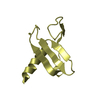
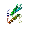
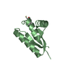
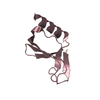
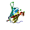
| Title | NMR solution structure of the Ca2+-Calmodulin C-terminal domain in a complex with a peptide (NSCaTE) from the L-type Voltage-Gated Calcium Channel alpha1C subunit | ||||||
 Components Components | Calmodulin | ||||||
 Keywords Keywords | METAL BINDING PROTEIN / NSCaTE | ||||||
| Function / homology |  Function and homology information Function and homology informationnegative regulation of calcium ion transmembrane transporter activity / : / establishment of protein localization to mitochondrial membrane / type 3 metabotropic glutamate receptor binding / regulation of synaptic vesicle endocytosis / negative regulation of high voltage-gated calcium channel activity / regulation of synaptic vesicle exocytosis / negative regulation of calcium ion export across plasma membrane / regulation of cardiac muscle cell action potential / response to corticosterone ...negative regulation of calcium ion transmembrane transporter activity / : / establishment of protein localization to mitochondrial membrane / type 3 metabotropic glutamate receptor binding / regulation of synaptic vesicle endocytosis / negative regulation of high voltage-gated calcium channel activity / regulation of synaptic vesicle exocytosis / negative regulation of calcium ion export across plasma membrane / regulation of cardiac muscle cell action potential / response to corticosterone / positive regulation of ryanodine-sensitive calcium-release channel activity / nitric-oxide synthase binding / regulation of cell communication by electrical coupling involved in cardiac conduction / negative regulation of peptidyl-threonine phosphorylation / protein phosphatase activator activity / positive regulation of cyclic-nucleotide phosphodiesterase activity / positive regulation of phosphoprotein phosphatase activity / adenylate cyclase binding / catalytic complex / detection of calcium ion / negative regulation of ryanodine-sensitive calcium-release channel activity / regulation of cardiac muscle contraction / positive regulation of DNA binding / calcium channel inhibitor activity / regulation of cardiac muscle contraction by regulation of the release of sequestered calcium ion / enzyme regulator activity / phosphatidylinositol 3-kinase binding / voltage-gated potassium channel complex / regulation of release of sequestered calcium ion into cytosol by sarcoplasmic reticulum / regulation of calcium-mediated signaling / positive regulation of protein dephosphorylation / titin binding / positive regulation of protein autophosphorylation / sperm midpiece / response to amphetamine / calcium channel complex / activation of adenylate cyclase activity / substantia nigra development / adenylate cyclase activator activity / protein serine/threonine kinase activator activity / regulation of heart rate / nitric-oxide synthase regulator activity / sarcomere / positive regulation of peptidyl-threonine phosphorylation / regulation of cytokinesis / positive regulation of nitric-oxide synthase activity / spindle microtubule / mitochondrial membrane / positive regulation of protein serine/threonine kinase activity / synaptic vesicle membrane / spindle pole / response to calcium ion / calcium-dependent protein binding / G2/M transition of mitotic cell cycle / disordered domain specific binding / myelin sheath / growth cone / vesicle / transmembrane transporter binding / G protein-coupled receptor signaling pathway / protein domain specific binding / centrosome / calcium ion binding / protein kinase binding / protein-containing complex / membrane / nucleus / plasma membrane / cytoplasmSimilarity search - Function | ||||||
| Biological species |  Homo sapiens (human) Homo sapiens (human) | ||||||
| Method | SOLUTION NMR / torsion angle dynamics, simulated annealing | ||||||
| Model details | lowest energy, model 1 | ||||||
 Authors Authors | Liu, Z. / Vogel, H.J. | ||||||
 Citation Citation | Journal: Front Mol Neurosci / Year: 2012 Title: Structural basis for the regulation of L-type voltage-gated calcium channels: interactions between the N-terminal cytoplasmic domain and Ca(2+)-calmodulin. Authors: Liu, Z. / Vogel, H.J. | ||||||
| History |
|
- Structure visualization
| Structure viewer | Molecule: MolmilJmol/JSmol |
|---|
- Downloads & links
-Download
| PDBx/mmCIF format | 2lqp.cif.gz | 403.2 KB | Display | PDBx/mmCIF format |
|---|---|---|---|---|
| PDB format | pdb2lqp.ent.gz | 339.2 KB | Display | PDB format |
| PDBx/mmJSON format | 2lqp.json.gz | Tree view | PDBx/mmJSON format | |
| Others |  Other downloads Other downloads |
-Validation report
| Arichive directory | https://data.pdbj.org/pub/pdb/validation_reports/lq/2lqpftp://data.pdbj.org/pub/pdb/validation_reports/lq/2lqp | HTTPS FTP |
|---|
-Related structure data


-Links
 PDBj
PDBj

- Assembly
| Deposited unit | 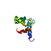
| |||||||||
|---|---|---|---|---|---|---|---|---|---|---|
| 1 |
| |||||||||
| NMR ensembles |
|
-Components
| #1: Protein | / CaM Mass: 8155.828 Da / Num. of mol.: 1 / Fragment: EF-hands 3 and 4 Source method: isolated from a genetically manipulated source Source: (gene. exp.) Homo sapiens (human)Gene: CALM1, CALM, CAM, CAM1, CALM2, CAM2, CAMB, CALM3, CALML2, CAM3, CAMC, CAMIII Production host:  Escherichia coli (E. coli) / References: UniProt: P62158, UniProt: P0DP24*PLUS Escherichia coli (E. coli) / References: UniProt: P62158, UniProt: P0DP24*PLUS |
|---|---|
| #2: Chemical |   Mass: 40.078 Da / Num. of mol.: 2 / Source method: obtained synthetically / Formula: Ca Mass: 40.078 Da / Num. of mol.: 2 / Source method: obtained synthetically / Formula: Ca |
-Experimental details
-Experiment
| Experiment | Method: SOLUTION NMR | ||||||||||||||||||||||||||||||||||||||||||||||||||||||||
|---|---|---|---|---|---|---|---|---|---|---|---|---|---|---|---|---|---|---|---|---|---|---|---|---|---|---|---|---|---|---|---|---|---|---|---|---|---|---|---|---|---|---|---|---|---|---|---|---|---|---|---|---|---|---|---|---|---|
| NMR experiment |
|
- Sample preparation
| Details | Contents: 20 mM TRIS, 90% H2O/10% D2O / Solvent system: 90% H2O/10% D2O |
|---|---|
| Sample | Conc.: 20 mM / Component: TRIS-1 |
| Sample conditions | Ionic strength: 100 / pH: 7.0 / Pressure: ambient / Temperature: 298 K |
-NMR measurement
| NMR spectrometer | Type: Bruker Avance / Manufacturer: Bruker / Model: AVANCE / Field strength: 500 MHz |
|---|
- Processing
| NMR software |
| ||||||||||||||||||||||||
|---|---|---|---|---|---|---|---|---|---|---|---|---|---|---|---|---|---|---|---|---|---|---|---|---|---|
| Refinement | Method: torsion angle dynamics, simulated annealing / Software ordinal: 1 | ||||||||||||||||||||||||
| NMR representative | Selection criteria: lowest energy | ||||||||||||||||||||||||
| NMR ensemble | Conformer selection criteria: structures with the lowest energy Conformers calculated total number: 200 / Conformers submitted total number: 19 |
