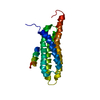
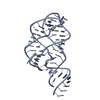
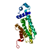
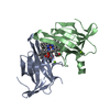
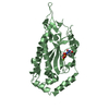
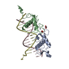
Mass: 22254.971 Da / Num. of mol.: 1 Fragment: FAT DOMAIN, UNP RESIDUES 916-1053, LINKER1, LD2, UNP RESIDUES 140-161, LINKER2, LD4, UNP RESIDUES 262-276 Source method: isolated from a genetically manipulated source Details: Fat domain of focal adhesion kinase with the Ld2 motif of paxillin tethered via linker1 GSGGSGSGGSGGSG which is then linked to Ld4 motif of paxillin via linker 2 GSGSGSGSGGSGGSGGSGGSGGSGGS Source: (gene. exp.) Gallus gallus, unidentified / Gene: PTK2, FAK, FAK1, PXN / Plasmid: pET28A / Production host: Escherichia coli (E. coli) / Strain (production host): BL21 References: UniProt: Q00944, UniProt: P49024, non-specific protein-tyrosine kinase
-
Experimental details
-
Experiment
Experiment
Method: SOLUTION NMR
NMR experiment
Conditions-ID
Experiment-ID
Solution-ID
Type
1
1
1
3D 1H-15N NOESY
1
2
1
3D 1H-13C NOESY
1
3
1
3D (H)CCH-TOCSY
1
4
1
3D (H)CCH-COSY
1
5
1
3D HNCA
1
6
1
3DCBCA(CO)NH
1
7
1
3D HNCO
1
8
1
3D HN(CA)CB
1
9
1
2D 1H-15N HSQC
1
10
1
2D 1H-13C HSQC
-
Sample preparation
Details
Contents: 1 mM [U-100% 13C; U-100% 15N] FOCAL ADHESION KINASE 1, LINKER, 22-MERIC PEPTIDE FROM PAXILLIN, LINKER, 15-MERIC PEPTIDE FROM PAXILLIN-1, 10 mM potassium phosphate-2, 0.1 % sodium azide-3, 90% H2O/10% D2O Solvent system: 90% H2O/10% D2O
Sample
Conc. (mg/ml)
Component
Isotopic labeling
Solution-ID
1mM
FOCAL ADHESION KINASE 1, LINKER, 22-MERIC PEPTIDE FROM PAXILLIN, LINKER, 15-MERIC PEPTIDE FROM PAXILLIN-1
[U-100% 13C; U-100% 15N]
1
10mM
potassium phosphate-2
1
0.1 %
sodium azide-3
1
Sample conditions
Ionic strength: 50 / pH: 6.5 / Pressure: AMBIENT Pa / Temperature: 310 K
-
NMR measurement
NMR spectrometer
Type
Manufacturer
Model
Field strength (MHz)
Spectrometer-ID
BRUKER AVANCE
Bruker
Avance
800
1
VARIAN INOVA
Varian
INOVA
600
2
-
Processing
NMR software
Name
Version
Developer
Classification
CYANA
Guntert, MumenthalerandWuthrich
refinement
CYANA
Guntert, MumenthalerandWuthrich
structuresolution
SPARKY
3
Goddard
structuresolution
NMRPIPE
Delaglio, Grzesiek, Vuister, Zhu, PfeiferandBax
structuresolution
MOLMOL
Koradi, BilleterandWuthrich
structuresolution
Refinement
Method: SIMULATED ANNEALING, TORSION ANGLE DYNAMICS / Software ordinal: 1 Details: THE SAMPLES USED ARE CONSTRUCTS IN WHICH ONE MOTIF IS TETHERED TO THE C-TERMINUS OF THE FAT DOMAIN VIA A GGSX LINKER AND THE OTHER LD MOTIF IS BOUND AS A FREE PEPTIDE. THE GGS LINKERS ARE ...Details: THE SAMPLES USED ARE CONSTRUCTS IN WHICH ONE MOTIF IS TETHERED TO THE C-TERMINUS OF THE FAT DOMAIN VIA A GGSX LINKER AND THE OTHER LD MOTIF IS BOUND AS A FREE PEPTIDE. THE GGS LINKERS ARE UNSTRUCTURED AND COMPARISON OF SPECTRA LEAD US TO BELIEVE THEY DO NOT IMPOSE ANY CONSTRAINT UPON HOW THE TETHERED LD MOTIFS BIND TO FAT. HOWEVER WHEN ANALYZING THE STRUCTURE IN CYANA, THE PRESENCE OF THE GGS LINKER IN THE SEQUENCE INTERFERS WITH CYANA'S INITIAL ALIGNMENT OF THE 20 LOWEST ENERGY STRUCTURES AND THUS CONFUSES RMSD AND TARGET FUNCTION CALCULATIONS. WE FOUND THAT USING PSEUDO LINKERS IN CYANA RESOLVED THIS PROBLEM WITHOUT CHANGING THE STRUCTURES IN QUESTION AND SO WE CHOSE TO SUBMIT THE STRUCTURES IN THAT WAY. 2RP6 REPRESENTS A COMPOSITE STRUCTURE USING CONSTRAINTS OBTAINED FROM THE 2RP7 AND 2RP9 DATA SETS. IT DOES NOT REPRESENT A NEW CONSTRUCT IN ITSELF, BUT MOST ACCURATELY REPRESENTS THE COMPLETE COMPLEX STRUCTURE BETWEEN PAXILLIN AND FOCAL ADHESION KINASE.
NMR representative
Selection criteria: lowest energy
NMR ensemble
Conformer selection criteria: target function / Conformers calculated total number: 500 / Conformers submitted total number: 20
+
About Yorodumi
-
News
-
Feb 9, 2022. New format data for meta-information of EMDB entries
New format data for meta-information of EMDB entries
Version 3 of the EMDB header file is now the official format.
The previous official version 1.9 will be removed from the archive.
In the structure databanks used in Yorodumi, some data are registered as the other names, "COVID-19 virus" and "2019-nCoV". Here are the details of the virus and the list of structure data.
Jan 31, 2019. EMDB accession codes are about to change! (news from PDBe EMDB page)
EMDB accession codes are about to change! (news from PDBe EMDB page)
The allocation of 4 digits for EMDB accession codes will soon come to an end. Whilst these codes will remain in use, new EMDB accession codes will include an additional digit and will expand incrementally as the available range of codes is exhausted. The current 4-digit format prefixed with “EMD-” (i.e. EMD-XXXX) will advance to a 5-digit format (i.e. EMD-XXXXX), and so on. It is currently estimated that the 4-digit codes will be depleted around Spring 2019, at which point the 5-digit format will come into force.
The EM Navigator/Yorodumi systems omit the EMD- prefix.
Related info.:Q: What is EMD? / ID/Accession-code notation in Yorodumi/EM Navigator
Yorodumi is a browser for structure data from EMDB, PDB, SASBDB, etc.
This page is also the successor to EM Navigator detail page, and also detail information page/front-end page for Omokage search.
The word "yorodu" (or yorozu) is an old Japanese word meaning "ten thousand". "mi" (miru) is to see.
Related info.:EMDB / PDB / SASBDB / Comparison of 3 databanks / Yorodumi Search / Aug 31, 2016. New EM Navigator & Yorodumi / Yorodumi Papers / Jmol/JSmol / Function and homology information / Changes in new EM Navigator and Yorodumi
 Movie
Movie Controller

 Yorodumi
 Open data
 Basic information
 Components
 PTK2
PTK2  Keywords
 Function and homology information
 Authors
 Citation
 Structure visualization
 Downloads & links
 Other downloads
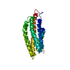


 PDBj
PDBj
 Assembly

 Sample preparation
 Processing