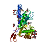
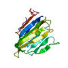
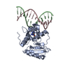
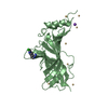
Entry Database : PDB / ID : 2jh7Title Crystal structure of Toxoplasma gondii micronemal protein 1 bound to 6'-sialyl-N-acetyllactosamine MICRONEMAL PROTEIN 1 Keywords / / / / Function / homology Biological species TOXOPLASMA GONDII (eukaryote)Method / / / Resolution : 2.07 Å Authors Blumenschein, T.M.A. / Friedrich, N. / Childs, R.A. / Saouros, S. / Carpenter, E.P. / Campanero-Rhodes, M.A. / Simpson, P. / Koutroukides, T. / Blackman, M.J. / Feizi, T. ...Blumenschein, T.M.A. / Friedrich, N. / Childs, R.A. / Saouros, S. / Carpenter, E.P. / Campanero-Rhodes, M.A. / Simpson, P. / Koutroukides, T. / Blackman, M.J. / Feizi, T. / Soldati-Favre, D. / Matthews, S. Journal : Embo J. / Year : 2007Title : Atomic Resolution Insight Into Host Cell Recognition by Toxoplasma Gondii.Authors: Blumenschein, T.M.A. / Friedrich, N. / Childs, R.A. / Saouros, S. / Carpenter, E.P. / Campanero-Rhodes, M.A. / Simpson, P. / Chai, W. / Koutroukides, T. / Blackman, M.J. / Feizi, T. / ... Authors : Blumenschein, T.M.A. / Friedrich, N. / Childs, R.A. / Saouros, S. / Carpenter, E.P. / Campanero-Rhodes, M.A. / Simpson, P. / Chai, W. / Koutroukides, T. / Blackman, M.J. / Feizi, T. / Soldati-Favre, D. / Matthews, S. History Deposition Feb 20, 2007 Deposition site / Processing site Revision 1.0 May 22, 2007 Provider / Type Revision 1.1 May 8, 2011 Group Revision 1.2 Jul 13, 2011 Group Revision 1.3 May 8, 2019 Group Data collection / Derived calculations ... Data collection / Derived calculations / Experimental preparation / Other Category database_PDB_rev / database_PDB_rev_record ... database_PDB_rev / database_PDB_rev_record / exptl_crystal_grow / pdbx_database_proc / pdbx_database_status / struct_conn Item / _pdbx_database_status.recvd_author_approval / _struct_conn.pdbx_leaving_atom_flagRevision 2.0 Jul 29, 2020 Group Atomic model / Data collection ... Atomic model / Data collection / Derived calculations / Other / Structure summary Category atom_site / chem_comp ... atom_site / chem_comp / entity / pdbx_branch_scheme / pdbx_chem_comp_identifier / pdbx_database_status / pdbx_entity_branch / pdbx_entity_branch_descriptor / pdbx_entity_branch_link / pdbx_entity_branch_list / pdbx_entity_nonpoly / pdbx_nonpoly_scheme / pdbx_struct_assembly_gen / pdbx_struct_special_symmetry / struct_asym / struct_conn / struct_site / struct_site_gen Item _atom_site.B_iso_or_equiv / _atom_site.Cartn_x ... _atom_site.B_iso_or_equiv / _atom_site.Cartn_x / _atom_site.Cartn_y / _atom_site.Cartn_z / _atom_site.auth_asym_id / _atom_site.auth_atom_id / _atom_site.auth_comp_id / _atom_site.auth_seq_id / _atom_site.label_asym_id / _atom_site.label_atom_id / _atom_site.label_comp_id / _atom_site.label_entity_id / _atom_site.type_symbol / _chem_comp.name / _chem_comp.type / _pdbx_database_status.status_code_sf / _pdbx_struct_assembly_gen.asym_id_list / _pdbx_struct_special_symmetry.label_asym_id / _struct_conn.pdbx_dist_value / _struct_conn.pdbx_leaving_atom_flag / _struct_conn.pdbx_value_order / _struct_conn.ptnr1_auth_asym_id / _struct_conn.ptnr1_auth_comp_id / _struct_conn.ptnr1_auth_seq_id / _struct_conn.ptnr1_label_atom_id / _struct_conn.ptnr1_label_comp_id / _struct_conn.ptnr2_auth_asym_id / _struct_conn.ptnr2_auth_comp_id / _struct_conn.ptnr2_auth_seq_id / _struct_conn.ptnr2_label_asym_id / _struct_conn.ptnr2_label_atom_id / _struct_conn.ptnr2_label_comp_id Description / Provider / Type Revision 2.1 Dec 13, 2023 Group Data collection / Database references ... Data collection / Database references / Derived calculations / Refinement description / Structure summary Category chem_comp / chem_comp_atom ... chem_comp / chem_comp_atom / chem_comp_bond / database_2 / pdbx_initial_refinement_model / struct_sheet Item _chem_comp.pdbx_synonyms / _database_2.pdbx_DOI ... _chem_comp.pdbx_synonyms / _database_2.pdbx_DOI / _database_2.pdbx_database_accession / _struct_sheet.number_strands
Show all Show less Remark 650 HELIX DETERMINATION METHOD: AUTHOR PROVIDED. Remark 700 SHEET DETERMINATION METHOD: AUTHOR PROVIDED.
 Movie
Movie Controller

 Yorodumi
 Open data
 Basic information
 Components
 Keywords
 CELL ADHESION / MIC1 /
  Function and homology information
 Authors
 Citation
 Structure visualization
 Downloads & links
 Other downloads
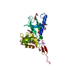


 PDBj
PDBj Assembly


 Mass: 59.044 Da / Num. of mol.: 13 / Source method: obtained synthetically / Formula: C2H3O2
 Mass: 35.453 Da / Num. of mol.: 2 / Source method: obtained synthetically / Formula: Cl
 Mass: 92.094 Da / Num. of mol.: 2 / Source method: obtained synthetically / Formula: C3H8O3
 Sample preparation
 / Beamline: PX14.1 / Wavelength: 1.488
  Processing