Group: Atomic model / Database references ...Atomic model / Database references / Derived calculations / Non-polymer description / Structure summary / Version format compliance
Revision 1.2
Nov 30, 2012
Group: Other
Revision 1.3
Mar 13, 2013
Group: Other
Revision 1.4
Aug 7, 2013
Group: Derived calculations / Other
Revision 1.5
Jun 28, 2017
Group: Data collection / Category: diffrn_source / Item: _diffrn_source.type
Remark 700
SHEET DETERMINATION METHOD: DSSP THE SHEETS PRESENTED AS "DB" IN EACH CHAIN ON SHEET RECORDS BELOW ... SHEET DETERMINATION METHOD: DSSP THE SHEETS PRESENTED AS "DB" IN EACH CHAIN ON SHEET RECORDS BELOW IS ACTUALLY AN 6-STRANDED BARREL THIS IS REPRESENTED BY A 7-STRANDED SHEET IN WHICH THE FIRST AND LAST STRANDS ARE IDENTICAL.
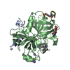
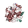
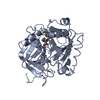
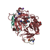
THROMBINLIGHTCHAIN / / COAGULATION FACTOR II / ALPHA THROMBIN
Mass: 4096.534 Da / Num. of mol.: 1 / Fragment: LIGHT CHAIN, RESIDUES 328-363 / Source method: isolated from a natural source / Details: LIGHT CHAIN HAS ONE DISULPHIDE BOND TO HEAVY CHAIN / Source: (natural) HOMO SAPIENS (human) / Tissue: BLOOD PLASMA / References: UniProt: P00734, thrombin
#3: Protein/peptide
HIRUDINIIIA / HIRUGEN
Mass: 1363.399 Da / Num. of mol.: 1 / Fragment: RESIDUES 55-64 / Source method: obtained synthetically / Details: RESIDUE 63 IS A SULFONATED TYROSINE / Source: (synth.) HIRUDO MEDICINALIS (medicinal leech) / References: UniProt: P28507
-
Protein , 1 types, 1 molecules D
#2: Protein
THROMBINHEAVYCHAIN / / COAGULATION FACTOR II / ALPHA THROMBIN
Mass: 29780.219 Da / Num. of mol.: 1 / Fragment: HEAVY CHAIN, RESIDUES 364-622 / Source method: isolated from a natural source / Details: LIGHT CHAIN HAS ONE DISULPHIDE BOND TO HEAVY CHAIN / Source: (natural) HOMO SAPIENS (human) / Tissue: BLOOD PLASMA / References: UniProt: P00734, thrombin
 Movie
Movie Controller


 Open data
 Basic information
 Components
 Keywords
 GLYCOPROTEIN /
  Function and homology information

 Authors
 Citation
 Structure visualization
 Downloads & links
 Other downloads
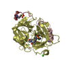


 PDBj
PDBj


 Assembly


 Mass: 40.078 Da / Num. of mol.: 1 / Source method: obtained synthetically / Formula: Ca
 Mass: 22.990 Da / Num. of mol.: 1 / Source method: obtained synthetically / Formula: Na
 Mass: 447.957 Da / Num. of mol.: 1 / Source method: obtained synthetically / Formula: C17H22ClN3O5S2
 Sample preparation
 Processing