Mass: 18.015 Da / Num. of mol.: 735 / Source method: isolated from a natural source / Formula: H2O
-
Experimental details
-
Experiment
Experiment
Method: X-RAY DIFFRACTION / Number of used crystals: 1
-
Sample preparation
Crystal
Density Matthews: 3.41 Å3/Da / Density % sol: 63.67 % Description: SELENIUM SITES WERE FOUND WITH MAD DATA IN SHELXD. PHASES WERE DETERMINED WITH SAD DATA IN PHASER
Protocol: SINGLE WAVELENGTH / Monochromatic (M) / Laue (L): M / Scattering type: x-ray
Radiation wavelength
Wavelength: 0.939272 Å / Relative weight: 1
Reflection
Resolution: 2.6→70 Å / Num. obs: 80455 / % possible obs: 99.9 % / Observed criterion σ(I): 2 / Redundancy: 8.7 % / Rmerge(I) obs: 0.14 / Net I/σ(I): 17.2
Reflection shell
Resolution: 2.6→2.69 Å / Redundancy: 8.6 % / Rmerge(I) obs: 0.87 / Mean I/σ(I) obs: 3 / % possible all: 99.8
-
Processing
Software
Name
Version
Classification
REFMAC
5.2.0019
refinement
DENZO
datareduction
SCALEPACK
datascaling
PHASER
phasing
Refinement
Method to determine structure: SAD / Resolution: 2.6→29.09 Å / Cor.coef. Fo:Fc: 0.943 / Cor.coef. Fo:Fc free: 0.9 / Cross valid method: THROUGHOUT / ESU R: 0.376 / ESU R Free: 0.276 / Stereochemistry target values: MAXIMUM LIKELIHOOD / Details: HYDROGENS HAVE BEEN ADDED IN THE RIDING POSITIONS.
Rfactor
Num. reflection
% reflection
Selection details
Rfree
0.25
4041
5 %
RANDOM
Rwork
0.186
-
-
-
obs
0.189
76341
99.9 %
-
Solvent computation
Ion probe radii: 0.8 Å / Shrinkage radii: 0.8 Å / VDW probe radii: 1.4 Å / Solvent model: MASK
 Movie
Movie Controller


 Open data
 Basic information
 Components
 Keywords
 OXIDOREDUCTASE /
  Function and homology information
 Authors
 Citation
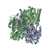
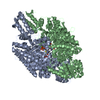
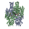
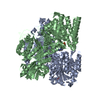
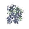
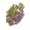
 Structure visualization
 Downloads & links
 Other downloads


 PDBj
PDBj


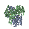
 Assembly
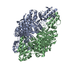

 Mass: 347.221 Da / Num. of mol.: 2 / Source method: obtained synthetically / Formula: C10H14N5O7P / Comment: AMP*YM
 Mass: 18.015 Da / Num. of mol.: 735 / Source method: isolated from a natural source / Formula: H2O
 Sample preparation
 / Beamline: ID14-4 / Wavelength: 0.939272
  Processing