 Movie
Movie Controller


+ Open data

- Basic information
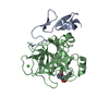
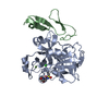
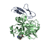
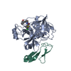
| Entry | Database: PDB / ID: 2j95 | ||||||
|---|---|---|---|---|---|---|---|
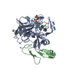
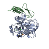
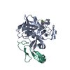
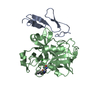
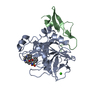
| Title | CRYSTAL STRUCTURE OF A HUMAN FACTOR XA INHIBITOR COMPLEX | ||||||
 Components Components |
| ||||||
 Keywords Keywords |  HYDROLASE / SERINE PROTEASE / EGF-LIKE DOMAIN / BLOOD COAGULATION / POLYMORPHISM / GLYCOPROTEIN / HYDROXYLATION / GAMMA- CARBOXYGLUTAMIC ACID / CALCIUM / ZYMOGEN / COMPLEX / PROTEASE HYDROLASE / SERINE PROTEASE / EGF-LIKE DOMAIN / BLOOD COAGULATION / POLYMORPHISM / GLYCOPROTEIN / HYDROXYLATION / GAMMA- CARBOXYGLUTAMIC ACID / CALCIUM / ZYMOGEN / COMPLEX / PROTEASE | ||||||
| Function / homology |  Function and homology informationcoagulation factor Xa / Defective factor IX causes thrombophilia / Defective cofactor function of FVIIIa variant / Defective F9 variant does not activate FX / Extrinsic Pathway of Fibrin Clot Formation / positive regulation of TOR signaling / Gamma-carboxylation of protein precursors / Transport of gamma-carboxylated protein precursors from the endoplasmic reticulum to the Golgi apparatus / Common Pathway of Fibrin Clot Formation / Removal of aminoterminal propeptides from gamma-carboxylated proteins ...coagulation factor Xa / Defective factor IX causes thrombophilia / Defective cofactor function of FVIIIa variant / Defective F9 variant does not activate FX / Extrinsic Pathway of Fibrin Clot Formation / positive regulation of TOR signaling / Gamma-carboxylation of protein precursors / Transport of gamma-carboxylated protein precursors from the endoplasmic reticulum to the Golgi apparatus / Common Pathway of Fibrin Clot Formation / Removal of aminoterminal propeptides from gamma-carboxylated proteins / Intrinsic Pathway of Fibrin Clot Formation / phospholipid binding / Golgi lumen / blood coagulation / positive regulation of cell migration / endoplasmic reticulum lumen / external side of plasma membrane / serine-type endopeptidase activity / calcium ion binding / proteolysis / extracellular space / extracellular region / plasma membrane Function and homology informationcoagulation factor Xa / Defective factor IX causes thrombophilia / Defective cofactor function of FVIIIa variant / Defective F9 variant does not activate FX / Extrinsic Pathway of Fibrin Clot Formation / positive regulation of TOR signaling / Gamma-carboxylation of protein precursors / Transport of gamma-carboxylated protein precursors from the endoplasmic reticulum to the Golgi apparatus / Common Pathway of Fibrin Clot Formation / Removal of aminoterminal propeptides from gamma-carboxylated proteins ...coagulation factor Xa / Defective factor IX causes thrombophilia / Defective cofactor function of FVIIIa variant / Defective F9 variant does not activate FX / Extrinsic Pathway of Fibrin Clot Formation / positive regulation of TOR signaling / Gamma-carboxylation of protein precursors / Transport of gamma-carboxylated protein precursors from the endoplasmic reticulum to the Golgi apparatus / Common Pathway of Fibrin Clot Formation / Removal of aminoterminal propeptides from gamma-carboxylated proteins / Intrinsic Pathway of Fibrin Clot Formation / phospholipid binding / Golgi lumen / blood coagulation / positive regulation of cell migration / endoplasmic reticulum lumen / external side of plasma membrane / serine-type endopeptidase activity / calcium ion binding / proteolysis / extracellular space / extracellular region / plasma membraneSimilarity search - Function | ||||||
| Biological species |  HOMO SAPIENS (human) HOMO SAPIENS (human) | ||||||
| Method | X-RAY DIFFRACTION / SYNCHROTRON / MOLECULAR REPLACEMENT / Resolution: 2.01 Å | ||||||
 Authors Authors | Chan, C. / Borthwick, A.D. / Brown, D. / Campbell, M. / Chaudry, L. / Chung, C.W. / Convery, M.A. / Hamblin, J.N. / Johnstone, L. / Kelly, H.A. ...Chan, C. / Borthwick, A.D. / Brown, D. / Campbell, M. / Chaudry, L. / Chung, C.W. / Convery, M.A. / Hamblin, J.N. / Johnstone, L. / Kelly, H.A. / Kleanthous, S. / Burns-Kurtis, C.L. / Patikis, A. / Patel, C. / Pateman, A.J. / Senger, S. / Shah, G.P. / Toomey, J.R. / Watson, N.S. / Weston, H.E. / Whitworth, C. / Young, R.J. / Zhou, P. | ||||||
 Citation Citation | Journal: J.Med.Chem. / Year: 2007 Title: Factor Xa Inhibitors: S1 Binding Interactions of a Series of N-{(3S)-1-[(1S)-1-Methyl-2-Morpholin-4-Yl-2-Oxoethyl]-2-Oxopyrrolidin-3-Yl}Sulfonamides. Authors: Chan, C. / Borthwick, A.D. / Brown, D. / Burns-Kurtis, C.L. / Campbell, M. / Chaudry, L. / Chung, C.W. / Convery, M.A. / Hamblin, J.N. / Johnstone, L. / Kelly, H.A. / Kleanthous, S. / ...Authors: Chan, C. / Borthwick, A.D. / Brown, D. / Burns-Kurtis, C.L. / Campbell, M. / Chaudry, L. / Chung, C.W. / Convery, M.A. / Hamblin, J.N. / Johnstone, L. / Kelly, H.A. / Kleanthous, S. / Patikis, A. / Patel, C. / Pateman, A.J. / Senger, S. / Shah, G.P. / Toomey, J.R. / Watson, N.S. / Weston, H.E. / Whitworth, C. / Young, R.J. / Zhou, P. | ||||||
| History |
|
- Structure visualization
| Structure viewer | Molecule: MolmilJmol/JSmol |
|---|
- Downloads & links
-Download
| PDBx/mmCIF format | 2j95.cif.gz | 79.9 KB | Display | PDBx/mmCIF format |
|---|---|---|---|---|
| PDB format | pdb2j95.ent.gz | 57.2 KB | Display | PDB format |
| PDBx/mmJSON format | 2j95.json.gz | Tree view | PDBx/mmJSON format | |
| Others |  Other downloads Other downloads |
-Validation report
| Arichive directory | https://data.pdbj.org/pub/pdb/validation_reports/j9/2j95ftp://data.pdbj.org/pub/pdb/validation_reports/j9/2j95 | HTTPS FTP |
|---|
-Related structure data
| Related structure data |  2j94C  4y76C  4y79C  1ezqS S: Starting model for refinement C: citing same article ( |
|---|---|
| Similar structure data |


-Links
 PDBj
PDBj


- Assembly
| Deposited unit | 
| ||||||||
|---|---|---|---|---|---|---|---|---|---|
| 1 |
| ||||||||
| Unit cell |
|
-Components
| #1: Protein | Mass: 28550.596 Da / Num. of mol.: 1 / Fragment: ACTIVATED DESGLA, RESIDUES 235-488 / Source method: isolated from a natural source Details: PURCHASED FROM ENZYME RESEARCH LABS. ISOLATED FROM HUMAN BLOOD Source: (natural) HOMO SAPIENS (human) / References: UniProt: P00742, coagulation factor Xa |
|---|---|
| #2: Protein | Mass: 15210.793 Da / Num. of mol.: 1 / Fragment: ACTIVATED DESGLA, RESIDUES 46-179 / Source method: isolated from a natural source Details: PURCHASED FROM ENZYME RESEARCH LABS. ISOLATED FROM HUMAN BLOOD Source: (natural) HOMO SAPIENS (human) / References: UniProt: P00742, coagulation factor Xa |
| #3: Chemical | ChemComp-GSX /   Mass: 504.043 Da / Num. of mol.: 1 / Source method: obtained synthetically / Formula: C19H22ClN3O5S3 Mass: 504.043 Da / Num. of mol.: 1 / Source method: obtained synthetically / Formula: C19H22ClN3O5S3 |
| #4: Chemical | ChemComp-CA /   Mass: 40.078 Da / Num. of mol.: 1 / Source method: obtained synthetically / Formula: Ca Mass: 40.078 Da / Num. of mol.: 1 / Source method: obtained synthetically / Formula: Ca |
| #5: Water | ChemComp-HOH / Water Mass: 18.015 Da / Num. of mol.: 168 / Source method: isolated from a natural source / Formula: H2O Mass: 18.015 Da / Num. of mol.: 168 / Source method: isolated from a natural source / Formula: H2O |
| Sequence details | SOME RESIDUES IN CHAIN ARE NOT SEEN IN THE ELECTRON DENSITY. SEQUENCE DATABASE RESIDUES 1-45 (THE ...SOME RESIDUES IN CHAIN ARE NOT SEEN IN THE ELECTRON DENSITY. SEQUENCE DATABASE RESIDUES 1-45 (THE GLA DOMAIN) WERE BIOCHEMICA |
-Experimental details
-Experiment
| Experiment | Method: X-RAY DIFFRACTION / Number of used crystals: 1 |
|---|
- Sample preparation
| Crystal | Density Matthews: 2.45 Å3/Da / Density % sol: 35.53 % |
|---|---|
| Crystal grow | pH: 5.85 / Details: 18% PEG 6K, 100MM MES PH5.75, 10MM CACL2, pH 5.85 |
-Data collection
| Diffraction | Mean temperature: 100 K |
|---|---|
| Diffraction source | Source: SYNCHROTRON / Site: SRS  / Beamline: PX9.6 / Wavelength: 0.87 / Beamline: PX9.6 / Wavelength: 0.87 |
| Detector | Type: ADSC CCD / Detector: CCD / Date: May 10, 2001 |
| Radiation | Protocol: SINGLE WAVELENGTH / Monochromatic (M) / Laue (L): M / Scattering type: x-ray |
| Radiation wavelength | Wavelength: 0.87 Å / Relative weight: 1 |
| Reflection | Resolution: 2→30 Å / Num. obs: 22752 / % possible obs: 99.5 % / Observed criterion σ(I): 0 / Redundancy: 4 % / Rmerge(I) obs: 0.05 |
| Reflection shell | Resolution: 2→2.07 Å / Rmerge(I) obs: 0.38 |
- Processing
| Software |
| ||||||||||||||||||||||||||||||||||||||||||||||||||||||||||||||||||||||||||||||||||||||||||||||||||||||||||||||||||||||||||||||||||||||||||||||||||||||||||||||||||||||||||||||||||||||
|---|---|---|---|---|---|---|---|---|---|---|---|---|---|---|---|---|---|---|---|---|---|---|---|---|---|---|---|---|---|---|---|---|---|---|---|---|---|---|---|---|---|---|---|---|---|---|---|---|---|---|---|---|---|---|---|---|---|---|---|---|---|---|---|---|---|---|---|---|---|---|---|---|---|---|---|---|---|---|---|---|---|---|---|---|---|---|---|---|---|---|---|---|---|---|---|---|---|---|---|---|---|---|---|---|---|---|---|---|---|---|---|---|---|---|---|---|---|---|---|---|---|---|---|---|---|---|---|---|---|---|---|---|---|---|---|---|---|---|---|---|---|---|---|---|---|---|---|---|---|---|---|---|---|---|---|---|---|---|---|---|---|---|---|---|---|---|---|---|---|---|---|---|---|---|---|---|---|---|---|---|---|---|---|
| Refinement | Method to determine structure: MOLECULAR REPLACEMENT Starting model: PDB ENTRY 1EZQ Resolution: 2.01→20 Å / Cor.coef. Fo:Fc: 0.954 / Cor.coef. Fo:Fc free: 0.932 / SU B: 4.524 / SU ML: 0.125 / Cross valid method: THROUGHOUT / ESU R: 0.18 / ESU R Free: 0.167 / Stereochemistry target values: MAXIMUM LIKELIHOOD / Details: HYDROGENS HAVE BEEN ADDED IN THE RIDING POSITIONS.
| ||||||||||||||||||||||||||||||||||||||||||||||||||||||||||||||||||||||||||||||||||||||||||||||||||||||||||||||||||||||||||||||||||||||||||||||||||||||||||||||||||||||||||||||||||||||
| Solvent computation | Ion probe radii: 0.8 Å / Shrinkage radii: 0.8 Å / VDW probe radii: 1.2 Å / Solvent model: BABINET MODEL WITH MASK | ||||||||||||||||||||||||||||||||||||||||||||||||||||||||||||||||||||||||||||||||||||||||||||||||||||||||||||||||||||||||||||||||||||||||||||||||||||||||||||||||||||||||||||||||||||||
| Displacement parameters | Biso mean: 36.95 Å2
| ||||||||||||||||||||||||||||||||||||||||||||||||||||||||||||||||||||||||||||||||||||||||||||||||||||||||||||||||||||||||||||||||||||||||||||||||||||||||||||||||||||||||||||||||||||||
| Refinement step | Cycle: LAST / Resolution: 2.01→20 Å
| ||||||||||||||||||||||||||||||||||||||||||||||||||||||||||||||||||||||||||||||||||||||||||||||||||||||||||||||||||||||||||||||||||||||||||||||||||||||||||||||||||||||||||||||||||||||
| Refine LS restraints |
|