 Movie
Movie Controller


+ Open data

- Basic information
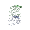
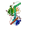
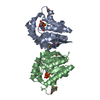
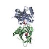
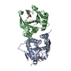
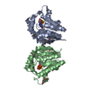
| Entry | Database: PDB / ID: 2j67 | ||||||
|---|---|---|---|---|---|---|---|
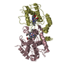
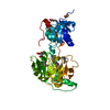
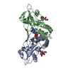
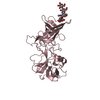
| Title | The TIR domain of human Toll-Like Receptor 10 (TLR10) | ||||||
 Components Components | TOLL LIKE RECEPTOR 10 Toll-like receptor Toll-like receptor | ||||||
 Keywords Keywords | RECEPTOR / TIR / IL-1 / TOLL / TLR10 / MEMBRANE / INFLAMMATORY RESPONSE / TOLL-LIKE RECEPTOR 10 / INNATE IMMUNITY / IMMUNE RESPONSE / LEUCINE-RICH REPEAT / GLYCOPROTEIN / TRANSMEMBRANE | ||||||
| Function / homology |  Function and homology information Function and homology informationtoll-like receptor 10 signaling pathway / Toll Like Receptor 10 (TLR10) Cascade / regulation of cytokine production => GO:0001817 / IRAK4 deficiency (TLR5) / MyD88 cascade initiated on plasma membrane / ADP-ribosyl cyclase/cyclic ADP-ribose hydrolase / NAD+ nucleotidase, cyclic ADP-ribose generating / NADP+ nucleosidase activity / MyD88-dependent toll-like receptor signaling pathway / toll-like receptor signaling pathway ...toll-like receptor 10 signaling pathway / Toll Like Receptor 10 (TLR10) Cascade / regulation of cytokine production => GO:0001817 / IRAK4 deficiency (TLR5) / MyD88 cascade initiated on plasma membrane / ADP-ribosyl cyclase/cyclic ADP-ribose hydrolase / NAD+ nucleotidase, cyclic ADP-ribose generating / NADP+ nucleosidase activity / MyD88-dependent toll-like receptor signaling pathway / toll-like receptor signaling pathway / plasma membrane => GO:0005886 / positive regulation of inflammatory response / transmembrane signaling receptor activity / signaling receptor activity / immune response / inflammatory response / innate immune response / membrane / identical protein binding / plasma membraneSimilarity search - Function | ||||||
| Biological species |  HOMO SAPIENS (human) HOMO SAPIENS (human) | ||||||
| Method | X-RAY DIFFRACTION / SYNCHROTRON / MOLECULAR REPLACEMENT / Resolution: 2.2 Å | ||||||
 Authors Authors | Stenmark, P. / Ogg, D. / Arrowsmith, C. / Berglund, H. / Busam, R. / Collins, R. / Edwards, A. / Ericsson, U.B. / Flodin, S. / Flores, A. ...Stenmark, P. / Ogg, D. / Arrowsmith, C. / Berglund, H. / Busam, R. / Collins, R. / Edwards, A. / Ericsson, U.B. / Flodin, S. / Flores, A. / Graslund, S. / Hammarstrom, M. / Hallberg, B.M. / Holmberg Schiavone, L. / Hogbom, M. / Johansson, I. / Karlberg, T. / Kotenyova, T. / Magnusdottir, A. / Nilsson, M.E. / Nilsson-Ehle, P. / Nyman, T. / Persson, C. / Sagemark, J. / Sundstrom, M. / Uppenberg, J. / Thorsell, A.G. / Van Den Berg, S. / Wallden, K. / Weigelt, J. / Welin, M. / Nordlund, P. | ||||||
 Citation Citation | Journal: J.Biol.Chem. / Year: 2008 Title: The Crystal Structure of the Human Toll-Like Receptor 10 Cytoplasmic Domain Reveals a Putative Signaling Dimer. Authors: Nyman, T. / Stenmark, P. / Flodin, S. / Johansson, I. / Hammarstrom, M. / Nordlund, P. | ||||||
| History |
|
- Structure visualization
| Structure viewer | Molecule: MolmilJmol/JSmol |
|---|
- Downloads & links
-Download
| PDBx/mmCIF format | 2j67.cif.gz | 74.7 KB | Display | PDBx/mmCIF format |
|---|---|---|---|---|
| PDB format | pdb2j67.ent.gz | 56.8 KB | Display | PDB format |
| PDBx/mmJSON format | 2j67.json.gz | Tree view | PDBx/mmJSON format | |
| Others |  Other downloads Other downloads |
-Validation report
| Arichive directory | https://data.pdbj.org/pub/pdb/validation_reports/j6/2j67ftp://data.pdbj.org/pub/pdb/validation_reports/j6/2j67 | HTTPS FTP |
|---|
-Related structure data
| Related structure data |  1fyvS S: Starting model for refinement |
|---|---|
| Similar structure data |
-Links
 PDBj
PDBj


- Assembly
| Deposited unit | 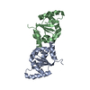
| ||||||||
|---|---|---|---|---|---|---|---|---|---|
| 1 |
| ||||||||
| Unit cell |
|
-Components
| #1: Protein | Toll-like receptor Mass: 21289.283 Da / Num. of mol.: 2 / Fragment: TIR DOMAIN, RESIDUES 622-776 Source method: isolated from a genetically manipulated source Source: (gene. exp.) HOMO SAPIENS (human) / Plasmid: PNIC-BSA4 / Production host:  ESCHERICHIA COLI (E. coli) / Strain (production host): BL21(DE3) / References: UniProt: Q9BXR5 ESCHERICHIA COLI (E. coli) / Strain (production host): BL21(DE3) / References: UniProt: Q9BXR5#2: Water | ChemComp-HOH / | Water Mass: 18.015 Da / Num. of mol.: 117 / Source method: isolated from a natural source / Formula: H2O Mass: 18.015 Da / Num. of mol.: 117 / Source method: isolated from a natural source / Formula: H2O |
|---|
-Experimental details
-Experiment
| Experiment | Method: X-RAY DIFFRACTION / Number of used crystals: 1 |
|---|
- Sample preparation
| Crystal | Density Matthews: 2.89 Å3/Da / Density % sol: 57.16 % |
|---|---|
| Crystal grow | Details: 0.2 M NASCN, 11% PEG 3350 |
-Data collection
| Diffraction | Mean temperature: 100 K |
|---|---|
| Diffraction source | Source: SYNCHROTRON / Site: BESSY  / Beamline: 14.1 / Wavelength: 0.95373 / Beamline: 14.1 / Wavelength: 0.95373 |
| Detector | Type: MARRESEARCH / Detector: CCD / Date: Aug 22, 2006 |
| Radiation | Protocol: SINGLE WAVELENGTH / Monochromatic (M) / Laue (L): M / Scattering type: x-ray |
| Radiation wavelength | Wavelength: 0.95373 Å / Relative weight: 1 |
| Reflection | Resolution: 2.2→23 Å / Num. obs: 19993 / % possible obs: 97.6 % / Observed criterion σ(I): 0 / Redundancy: 3.8 % / Rmerge(I) obs: 0.06 / Net I/σ(I): 13.8 |
| Reflection shell | Resolution: 2.2→2.3 Å / Redundancy: 3.7 % / Rmerge(I) obs: 0.4 / Mean I/σ(I) obs: 3.5 / % possible all: 92 |
- Processing
| Software |
| ||||||||||||||||||||||||||||||||||||||||||||||||||||||||||||||||||||||||||||||||||||||||||||||||||||||||||||||||||||||||||||||||||||||||||||||||||||||||||||||||||||||||||||||||||||||
|---|---|---|---|---|---|---|---|---|---|---|---|---|---|---|---|---|---|---|---|---|---|---|---|---|---|---|---|---|---|---|---|---|---|---|---|---|---|---|---|---|---|---|---|---|---|---|---|---|---|---|---|---|---|---|---|---|---|---|---|---|---|---|---|---|---|---|---|---|---|---|---|---|---|---|---|---|---|---|---|---|---|---|---|---|---|---|---|---|---|---|---|---|---|---|---|---|---|---|---|---|---|---|---|---|---|---|---|---|---|---|---|---|---|---|---|---|---|---|---|---|---|---|---|---|---|---|---|---|---|---|---|---|---|---|---|---|---|---|---|---|---|---|---|---|---|---|---|---|---|---|---|---|---|---|---|---|---|---|---|---|---|---|---|---|---|---|---|---|---|---|---|---|---|---|---|---|---|---|---|---|---|---|---|
| Refinement | Method to determine structure: MOLECULAR REPLACEMENT Starting model: PDB ENTRY 1FYV Resolution: 2.2→23.43 Å / Cor.coef. Fo:Fc: 0.933 / Cor.coef. Fo:Fc free: 0.903 / SU B: 12.12 / SU ML: 0.16 / TLS residual ADP flag: LIKELY RESIDUAL / Cross valid method: THROUGHOUT / ESU R: 0.252 / ESU R Free: 0.217 / Stereochemistry target values: MAXIMUM LIKELIHOOD / Details: HYDROGENS HAVE BEEN ADDED IN THE RIDING POSITIONS.
| ||||||||||||||||||||||||||||||||||||||||||||||||||||||||||||||||||||||||||||||||||||||||||||||||||||||||||||||||||||||||||||||||||||||||||||||||||||||||||||||||||||||||||||||||||||||
| Solvent computation | Ion probe radii: 0.8 Å / Shrinkage radii: 0.8 Å / VDW probe radii: 1.4 Å / Solvent model: BABINET MODEL WITH MASK | ||||||||||||||||||||||||||||||||||||||||||||||||||||||||||||||||||||||||||||||||||||||||||||||||||||||||||||||||||||||||||||||||||||||||||||||||||||||||||||||||||||||||||||||||||||||
| Displacement parameters | Biso mean: 28.04 Å2
| ||||||||||||||||||||||||||||||||||||||||||||||||||||||||||||||||||||||||||||||||||||||||||||||||||||||||||||||||||||||||||||||||||||||||||||||||||||||||||||||||||||||||||||||||||||||
| Refinement step | Cycle: LAST / Resolution: 2.2→23.43 Å
| ||||||||||||||||||||||||||||||||||||||||||||||||||||||||||||||||||||||||||||||||||||||||||||||||||||||||||||||||||||||||||||||||||||||||||||||||||||||||||||||||||||||||||||||||||||||
| Refine LS restraints |
|