 Movie
Movie Controller


+ Open data

- Basic information
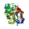
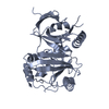
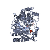
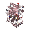
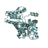
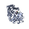
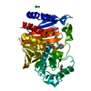
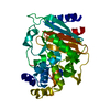
| Entry | Database: PDB / ID: 2j63 | ||||||
|---|---|---|---|---|---|---|---|
| Title | Crystal structure of AP endonuclease LMAP from Leishmania major | ||||||
 Components Components | AP-ENDONUCLEASE | ||||||
 Keywords Keywords |  LYASE / LEISHMANIA / ENDONUCLEASE / BASE EXCISION REPAIR LYASE / LEISHMANIA / ENDONUCLEASE / BASE EXCISION REPAIR | ||||||
| Function / homology |  Function and homology information Function and homology informationdouble-stranded DNA 3'-5' DNA exonuclease activity / phosphodiesterase I activity / DNA-(apurinic or apyrimidinic site) endonuclease activity / base-excision repair / endonuclease activity / Hydrolases; Acting on ester bonds / lyase activity / DNA repair / DNA binding / metal ion binding / nucleusSimilarity search - Function | ||||||
| Biological species |  LEISHMANIA MAJOR (eukaryote) LEISHMANIA MAJOR (eukaryote) | ||||||
| Method | X-RAY DIFFRACTION / SYNCHROTRON / MOLECULAR REPLACEMENT / Resolution: 2.48 Å | ||||||
 Authors Authors | Vidal, A.E. / Harkiolaki, M. / Gallego, C. / Castillo-Acosta, V.M. / Ruiz-Perez, L.M. / Wilson, K.S. / Gonzalez-Pacanowska, D. | ||||||
 Citation Citation | Journal: J.Mol.Biol. / Year: 2007 Title: Crystal Structure and DNA Repair Activities of the Ap Endonuclease from Leishmania Major. Authors: Vidal, A.E. / Harkiolaki, M. / Gallego, C. / Castillo-Acosta, V.M. / Ruiz-Perez, L.M. / Wilson, K. / Gonzalez-Pacanowska, D. | ||||||
| History |
|
- Structure visualization
| Structure viewer | Molecule: MolmilJmol/JSmol |
|---|
- Downloads & links
-Download
| PDBx/mmCIF format | 2j63.cif.gz | 147.8 KB | Display | PDBx/mmCIF format |
|---|---|---|---|---|
| PDB format | pdb2j63.ent.gz | 114.3 KB | Display | PDB format |
| PDBx/mmJSON format | 2j63.json.gz | Tree view | PDBx/mmJSON format | |
| Others |  Other downloads Other downloads |
-Validation report
| Arichive directory | https://data.pdbj.org/pub/pdb/validation_reports/j6/2j63ftp://data.pdbj.org/pub/pdb/validation_reports/j6/2j63 | HTTPS FTP |
|---|
-Related structure data
| Related structure data |  1de9S S: Starting model for refinement |
|---|---|
| Similar structure data |

-Links
 PDBj
PDBj

- Assembly
| Deposited unit | 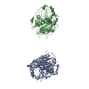
| ||||||||||||||||||
|---|---|---|---|---|---|---|---|---|---|---|---|---|---|---|---|---|---|---|---|
| 1 | 
| ||||||||||||||||||
| 2 | 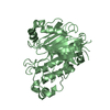
| ||||||||||||||||||
| Unit cell |
| ||||||||||||||||||
| Noncrystallographic symmetry (NCS) | NCS domain:
NCS domain segments: Component-ID: 1 / Ens-ID: 1 / Beg auth comp-ID: ILE / Beg label comp-ID: ILE / End auth comp-ID: HIS / End label comp-ID: HIS / Refine code: 2 / Auth seq-ID: 90 - 446 / Label seq-ID: 110 - 466
|
-Components
| #1: Protein | Mass: 50866.961 Da / Num. of mol.: 2 Source method: isolated from a genetically manipulated source Source: (gene. exp.) LEISHMANIA MAJOR (eukaryote) / Strain: 252 / Description: ISOLATED IN IRAN AND PROVIDED BY S. MESHNIK / Plasmid: PET-28A / Production host:  ESCHERICHIA COLI (E. coli) / Strain (production host): BL21(DE3) ESCHERICHIA COLI (E. coli) / Strain (production host): BL21(DE3)References: UniProt: O15922, DNA-(apurinic or apyrimidinic site) lyase#2: Water | ChemComp-HOH / | Water Mass: 18.015 Da / Num. of mol.: 135 / Source method: isolated from a natural source / Formula: H2O Mass: 18.015 Da / Num. of mol.: 135 / Source method: isolated from a natural source / Formula: H2OSequence details | POLY-HISTIDINE N-TERMINUS ADDED FOR PURIFICATI | |
|---|
-Experimental details
-Experiment
| Experiment | Method: X-RAY DIFFRACTION / Number of used crystals: 1 |
|---|
- Sample preparation
| Crystal | Density Matthews: 1.99 Å3/Da / Density % sol: 38.13 % / Description: NONE |
|---|---|
| Crystal grow | Temperature: 290 K / Method: vapor diffusion, hanging drop Details: HAMPTON SCREEN I CONDITION 42: 20% PEG 8000, 50MM POTASSIUM PHOSPHATE. HANGING DROPS AT 17C |
-Data collection
| Diffraction | Mean temperature: 100 K |
|---|---|
| Diffraction source | Source: SYNCHROTRON / Site: ESRF  / Beamline: ID14-2 / Wavelength: 0.933 / Beamline: ID14-2 / Wavelength: 0.933 |
| Detector | Type: ADSC QUANTUM 4 / Detector: CCD / Date: Oct 16, 2001 / Details: MIRRORS |
| Radiation | Monochromator: DIAMOND (111) / Protocol: SINGLE WAVELENGTH / Monochromatic (M) / Laue (L): M / Scattering type: x-ray |
| Radiation wavelength | Wavelength: 0.933 Å / Relative weight: 1 |
| Reflection | Resolution: 2.5→30.6 Å / Num. obs: 27305 / % possible obs: 99.9 % / Observed criterion σ(I): 3 / Redundancy: 30 % / Rmerge(I) obs: 0.05 / Net I/σ(I): 17.5 |
| Reflection shell | Resolution: 2.5→2.54 Å / Redundancy: 37.3 % / Rmerge(I) obs: 0.24 / Mean I/σ(I) obs: 2.6 / % possible all: 99.8 |
- Processing
| Software |
| ||||||||||||||||||||||||||||||||||||||||||||||||||||||||||||||||||||||||||||||||||||||||||||||||||||||||||||||||||||||||||||||||||||||||||||||||||||||||||||||||||||||||||||||||||||||
|---|---|---|---|---|---|---|---|---|---|---|---|---|---|---|---|---|---|---|---|---|---|---|---|---|---|---|---|---|---|---|---|---|---|---|---|---|---|---|---|---|---|---|---|---|---|---|---|---|---|---|---|---|---|---|---|---|---|---|---|---|---|---|---|---|---|---|---|---|---|---|---|---|---|---|---|---|---|---|---|---|---|---|---|---|---|---|---|---|---|---|---|---|---|---|---|---|---|---|---|---|---|---|---|---|---|---|---|---|---|---|---|---|---|---|---|---|---|---|---|---|---|---|---|---|---|---|---|---|---|---|---|---|---|---|---|---|---|---|---|---|---|---|---|---|---|---|---|---|---|---|---|---|---|---|---|---|---|---|---|---|---|---|---|---|---|---|---|---|---|---|---|---|---|---|---|---|---|---|---|---|---|---|---|
| Refinement | Method to determine structure: MOLECULAR REPLACEMENT Starting model: PDB ENTRY 1DE9 Resolution: 2.48→105.41 Å / Cor.coef. Fo:Fc: 0.947 / Cor.coef. Fo:Fc free: 0.916 / SU B: 20.974 / SU ML: 0.231 / TLS residual ADP flag: LIKELY RESIDUAL / Cross valid method: THROUGHOUT / ESU R: 0.59 / ESU R Free: 0.286 / Stereochemistry target values: MAXIMUM LIKELIHOOD / Details: HYDROGENS HAVE BEEN ADDED IN THE RIDING POSITIONS.
| ||||||||||||||||||||||||||||||||||||||||||||||||||||||||||||||||||||||||||||||||||||||||||||||||||||||||||||||||||||||||||||||||||||||||||||||||||||||||||||||||||||||||||||||||||||||
| Solvent computation | Ion probe radii: 0.8 Å / Shrinkage radii: 0.8 Å / VDW probe radii: 1.2 Å / Solvent model: MASK | ||||||||||||||||||||||||||||||||||||||||||||||||||||||||||||||||||||||||||||||||||||||||||||||||||||||||||||||||||||||||||||||||||||||||||||||||||||||||||||||||||||||||||||||||||||||
| Displacement parameters | Biso mean: 44.33 Å2
| ||||||||||||||||||||||||||||||||||||||||||||||||||||||||||||||||||||||||||||||||||||||||||||||||||||||||||||||||||||||||||||||||||||||||||||||||||||||||||||||||||||||||||||||||||||||
| Refinement step | Cycle: LAST / Resolution: 2.48→105.41 Å
| ||||||||||||||||||||||||||||||||||||||||||||||||||||||||||||||||||||||||||||||||||||||||||||||||||||||||||||||||||||||||||||||||||||||||||||||||||||||||||||||||||||||||||||||||||||||
| Refine LS restraints |
|