 Movie
Movie Controller


+ Open data

- Basic information
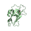
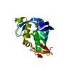
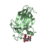
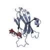
| Entry | Database: PDB / ID: 2j1l | ||||||
|---|---|---|---|---|---|---|---|
| Title | Crystal Structure of Human Rho-related GTP-binding protein RhoD | ||||||
 Components Components | RHO-RELATED GTP-BINDING PROTEIN RHOD | ||||||
 Keywords Keywords |  HYDROLASE / GTPASE / MEMBRANE / GTP-BINDING / PRENYLATION / NUCLEOTIDE-BINDING / METHYLATION / LIPOPROTEIN / ENDOSOME DYNAMICS HYDROLASE / GTPASE / MEMBRANE / GTP-BINDING / PRENYLATION / NUCLEOTIDE-BINDING / METHYLATION / LIPOPROTEIN / ENDOSOME DYNAMICS | ||||||
| Function / homology |  Function and homology informationfocal adhesion assembly / RHOD GTPase cycle / regulation of small GTPase mediated signal transduction / lamellipodium assembly / actin filament bundle assembly / Rho protein signal transduction / positive regulation of cell adhesion / guanyl-nucleotide exchange factor activity / actin filament organization / RHO GTPases Activate Formins ...focal adhesion assembly / RHOD GTPase cycle / regulation of small GTPase mediated signal transduction / lamellipodium assembly / actin filament bundle assembly / Rho protein signal transduction / positive regulation of cell adhesion / guanyl-nucleotide exchange factor activity / actin filament organization / RHO GTPases Activate Formins / cell migration / mitochondrial outer membrane / early endosome / endosome membrane / positive regulation of cell migration / Golgi membrane / GTPase activity / GTP binding / protein kinase binding / signal transduction / plasma membrane / cytosol Function and homology informationfocal adhesion assembly / RHOD GTPase cycle / regulation of small GTPase mediated signal transduction / lamellipodium assembly / actin filament bundle assembly / Rho protein signal transduction / positive regulation of cell adhesion / guanyl-nucleotide exchange factor activity / actin filament organization / RHO GTPases Activate Formins ...focal adhesion assembly / RHOD GTPase cycle / regulation of small GTPase mediated signal transduction / lamellipodium assembly / actin filament bundle assembly / Rho protein signal transduction / positive regulation of cell adhesion / guanyl-nucleotide exchange factor activity / actin filament organization / RHO GTPases Activate Formins / cell migration / mitochondrial outer membrane / early endosome / endosome membrane / positive regulation of cell migration / Golgi membrane / GTPase activity / GTP binding / protein kinase binding / signal transduction / plasma membrane / cytosolSimilarity search - Function | ||||||
| Biological species |  HOMO SAPIENS (human) HOMO SAPIENS (human) | ||||||
| Method | X-RAY DIFFRACTION / SYNCHROTRON / MOLECULAR REPLACEMENT / Resolution: 2.5 Å | ||||||
 Authors Authors | Pike, A.C.W. / Johansson, C. / Gileadi, C. / Niesen, F.H. / Sobott, F. / Schoch, G. / Elkins, J. / Smee, C. / Gorrec, F. / Watt, S. ...Pike, A.C.W. / Johansson, C. / Gileadi, C. / Niesen, F.H. / Sobott, F. / Schoch, G. / Elkins, J. / Smee, C. / Gorrec, F. / Watt, S. / Bray, J. / Turnbull, A.P. / von Delft, F. / Arrowsmith, C. / Edwards, A. / Weigelt, J. / Sundstrom, M. / Doyle, D. | ||||||
 Citation Citation | Journal: To be Published Title: Crystal Structure of Human Rho-Related GTP-Binding Protein Rhod Authors: Pike, A.C.W. / Johansson, C. / Gileadi, C. / Niesen, F.H. / Sobott, F. / Schoch, G. / Elkins, J. / Smee, C. / Gorrec, F. / Watt, S. / Bray, J. / Turnbull, A.P. / von Delft, F. / Arrowsmith, ...Authors: Pike, A.C.W. / Johansson, C. / Gileadi, C. / Niesen, F.H. / Sobott, F. / Schoch, G. / Elkins, J. / Smee, C. / Gorrec, F. / Watt, S. / Bray, J. / Turnbull, A.P. / von Delft, F. / Arrowsmith, C. / Edwards, A. / Weigelt, J. / Sundstrom, M. / Doyle, D. | ||||||
| History |
|
- Structure visualization
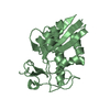
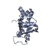
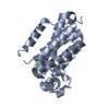
| Structure viewer | Molecule: MolmilJmol/JSmol |
|---|
- Downloads & links
-Download
| PDBx/mmCIF format | 2j1l.cif.gz | 46.9 KB | Display | PDBx/mmCIF format |
|---|---|---|---|---|
| PDB format | pdb2j1l.ent.gz | 30.9 KB | Display | PDB format |
| PDBx/mmJSON format | 2j1l.json.gz | Tree view | PDBx/mmJSON format | |
| Others |  Other downloads Other downloads |
-Validation report
| Arichive directory | https://data.pdbj.org/pub/pdb/validation_reports/j1/2j1lftp://data.pdbj.org/pub/pdb/validation_reports/j1/2j1l | HTTPS FTP |
|---|
-Related structure data
| Related structure data | 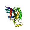 1mh1S S: Starting model for refinement |
|---|---|
| Similar structure data |


-Links
 PDBj
PDBj


- Assembly
| Deposited unit | 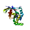
| ||||||||
|---|---|---|---|---|---|---|---|---|---|
| 1 |
| ||||||||
| Unit cell |
|
-Components
| #1: Protein | Mass: 24016.293 Da / Num. of mol.: 1 Source method: isolated from a genetically manipulated source Source: (gene. exp.) HOMO SAPIENS (human) / Description: MGC / Plasmid: PNIC-BSA4 / Production host:  ESCHERICHIA COLI (E. coli) / Strain (production host): BL21(DE3) / References: UniProt: O00212, small monomeric GTPase ESCHERICHIA COLI (E. coli) / Strain (production host): BL21(DE3) / References: UniProt: O00212, small monomeric GTPase |
|---|---|
| #2: Chemical | ChemComp-GDP / Guanosine diphosphate  Type: RNA linking / Mass: 443.201 Da / Num. of mol.: 1 / Source method: obtained synthetically / Formula: C10H15N5O11P2 / Comment: GDP, energy-carrying molecule*YM Type: RNA linking / Mass: 443.201 Da / Num. of mol.: 1 / Source method: obtained synthetically / Formula: C10H15N5O11P2 / Comment: GDP, energy-carrying molecule*YM |
| #3: Chemical | ChemComp-MG /   Mass: 24.305 Da / Num. of mol.: 1 / Source method: obtained synthetically / Formula: Mg Mass: 24.305 Da / Num. of mol.: 1 / Source method: obtained synthetically / Formula: Mg |
| #4: Water | ChemComp-HOH / Water Mass: 18.015 Da / Num. of mol.: 8 / Source method: isolated from a natural source / Formula: H2O Mass: 18.015 Da / Num. of mol.: 8 / Source method: isolated from a natural source / Formula: H2O |
-Experimental details
-Experiment
| Experiment | Method: X-RAY DIFFRACTION / Number of used crystals: 1 |
|---|
- Sample preparation
| Crystal | Density Matthews: 2.79 Å3/Da / Density % sol: 56 % |
|---|---|
| Crystal grow | pH: 6 Details: 19% PEG6K, 0.4M NH4CL, 15% ETG, 0.05M MES PH6.0, pH 6.00 |
-Data collection
| Diffraction | Mean temperature: 100 K |
|---|---|
| Diffraction source | Source: SYNCHROTRON / Site: SLS  / Beamline: X10SA / Wavelength: 0.979 / Beamline: X10SA / Wavelength: 0.979 |
| Detector | Type: MARRESEARCH / Detector: CCD / Date: May 6, 2006 |
| Radiation | Protocol: SINGLE WAVELENGTH / Monochromatic (M) / Laue (L): M / Scattering type: x-ray |
| Radiation wavelength | Wavelength: 0.979 Å / Relative weight: 1 |
| Reflection | Resolution: 2.5→50.6 Å / Num. obs: 9805 / % possible obs: 100 % / Observed criterion σ(I): 0 / Redundancy: 8.1 % / Biso Wilson estimate: 82.9 Å2 / Rmerge(I) obs: 0.09 / Net I/σ(I): 16.8 |
| Reflection shell | Resolution: 2.5→2.62 Å / Redundancy: 5.3 % / Rmerge(I) obs: 0.66 / Mean I/σ(I) obs: 2.3 / % possible all: 100 |
- Processing
| Software |
| ||||||||||||||||||||||||||||||||||||||||||||||||||||||||||||||||||||||||||||||||||||||||||||||||||||||||||||||||||||||||||||||||||||||||||||||||||||||||||||||||||||||||||||||||||||||
|---|---|---|---|---|---|---|---|---|---|---|---|---|---|---|---|---|---|---|---|---|---|---|---|---|---|---|---|---|---|---|---|---|---|---|---|---|---|---|---|---|---|---|---|---|---|---|---|---|---|---|---|---|---|---|---|---|---|---|---|---|---|---|---|---|---|---|---|---|---|---|---|---|---|---|---|---|---|---|---|---|---|---|---|---|---|---|---|---|---|---|---|---|---|---|---|---|---|---|---|---|---|---|---|---|---|---|---|---|---|---|---|---|---|---|---|---|---|---|---|---|---|---|---|---|---|---|---|---|---|---|---|---|---|---|---|---|---|---|---|---|---|---|---|---|---|---|---|---|---|---|---|---|---|---|---|---|---|---|---|---|---|---|---|---|---|---|---|---|---|---|---|---|---|---|---|---|---|---|---|---|---|---|---|
| Refinement | Method to determine structure: MOLECULAR REPLACEMENT Starting model: PDB ENTRY 1MH1 Resolution: 2.5→50 Å / Cor.coef. Fo:Fc: 0.955 / Cor.coef. Fo:Fc free: 0.947 / SU B: 17.082 / SU ML: 0.188 / TLS residual ADP flag: LIKELY RESIDUAL / Cross valid method: THROUGHOUT / ESU R: 0.285 / ESU R Free: 0.221 / Stereochemistry target values: MAXIMUM LIKELIHOOD Details: HYDROGENS HAVE BEEN ADDED IN THE RIDING POSITIONS. ELECTRON DENSITY SUGGESTS HIGH LIKELIHOOD OF A DOMAIN SWAP. IN THE CRYSTAL, THE AMINO TERMINAL RESIDUES 15- 39 APPEAR TO BE EXCHANGED WITH ...Details: HYDROGENS HAVE BEEN ADDED IN THE RIDING POSITIONS. ELECTRON DENSITY SUGGESTS HIGH LIKELIHOOD OF A DOMAIN SWAP. IN THE CRYSTAL, THE AMINO TERMINAL RESIDUES 15- 39 APPEAR TO BE EXCHANGED WITH AN ADJACENT MOLECULE. HOWEVER THE INTERVENING 'SWITCH 1' REGION BETWEEN RESIDUES 39 AND 47 IS DISORDERED. CONSEQUENTLY THE COMPACT FORM OF THE MOLECULE IS PRESENTED IN THIS ENTRY AS THE DOMAIN SWAP CANNOT BE CONFIRMED UNAMBIGUOUSLY. MASS SPECTROMETRIC ANALYSIS OF THE CRYSTALS CONFIRM THAT THE MOLECULE IS INTACT.
| ||||||||||||||||||||||||||||||||||||||||||||||||||||||||||||||||||||||||||||||||||||||||||||||||||||||||||||||||||||||||||||||||||||||||||||||||||||||||||||||||||||||||||||||||||||||
| Solvent computation | Ion probe radii: 0.8 Å / Shrinkage radii: 0.8 Å / VDW probe radii: 1.4 Å / Solvent model: MASK | ||||||||||||||||||||||||||||||||||||||||||||||||||||||||||||||||||||||||||||||||||||||||||||||||||||||||||||||||||||||||||||||||||||||||||||||||||||||||||||||||||||||||||||||||||||||
| Displacement parameters | Biso mean: 51.86 Å2
| ||||||||||||||||||||||||||||||||||||||||||||||||||||||||||||||||||||||||||||||||||||||||||||||||||||||||||||||||||||||||||||||||||||||||||||||||||||||||||||||||||||||||||||||||||||||
| Refinement step | Cycle: LAST / Resolution: 2.5→50 Å
| ||||||||||||||||||||||||||||||||||||||||||||||||||||||||||||||||||||||||||||||||||||||||||||||||||||||||||||||||||||||||||||||||||||||||||||||||||||||||||||||||||||||||||||||||||||||
| Refine LS restraints |
|