 Movie
Movie Controller


+ Open data

- Basic information
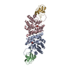
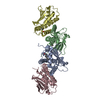
| Entry | Database: PDB / ID: 2htm | ||||||
|---|---|---|---|---|---|---|---|
| Title | Crystal structure of TTHA0676 from Thermus thermophilus HB8 | ||||||
 Components Components |
| ||||||
 Keywords Keywords |  BIOSYNTHETIC PROTEIN / thiamin biosynthesis / ThiG / Thermus thermophilus HB8 / Structural Genomics / NPPSFA / National Project on Protein Structural and Functional Analyses / RIKEN Structural Genomics/Proteomics Initiative / RSGI BIOSYNTHETIC PROTEIN / thiamin biosynthesis / ThiG / Thermus thermophilus HB8 / Structural Genomics / NPPSFA / National Project on Protein Structural and Functional Analyses / RIKEN Structural Genomics/Proteomics Initiative / RSGI | ||||||
| Function / homology |  Function and homology informationthiazole synthase / sulfurtransferase activity / thiamine diphosphate biosynthetic process / thiamine biosynthetic process / nucleotide binding / cytoplasm Function and homology informationthiazole synthase / sulfurtransferase activity / thiamine diphosphate biosynthetic process / thiamine biosynthetic process / nucleotide binding / cytoplasmSimilarity search - Function | ||||||
| Biological species |   Thermus thermophilus (bacteria) Thermus thermophilus (bacteria) | ||||||
| Method | X-RAY DIFFRACTION / SYNCHROTRON / MOLECULAR REPLACEMENT / Resolution: 2.3 Å | ||||||
 Authors Authors | Sugahara, M. / Kunishima, N. / RIKEN Structural Genomics/Proteomics Initiative (RSGI) | ||||||
 Citation Citation | Journal: To be Published Title: Crystal structure of TTHA0676 from Thermus thermophilus HB8 Authors: Sugahara, M. / Kunishima, N. | ||||||
| History |
|
- Structure visualization
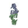
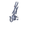
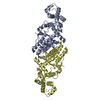
| Structure viewer | Molecule: MolmilJmol/JSmol |
|---|
- Downloads & links
-Download
| PDBx/mmCIF format | 2htm.cif.gz | 237 KB | Display | PDBx/mmCIF format |
|---|---|---|---|---|
| PDB format | pdb2htm.ent.gz | 190.7 KB | Display | PDB format |
| PDBx/mmJSON format | 2htm.json.gz | Tree view | PDBx/mmJSON format | |
| Others |  Other downloads Other downloads |
-Validation report
| Arichive directory | https://data.pdbj.org/pub/pdb/validation_reports/ht/2htmftp://data.pdbj.org/pub/pdb/validation_reports/ht/2htm | HTTPS FTP |
|---|
-Related structure data
| Related structure data |  1wv2S S: Starting model for refinement |
|---|---|
| Similar structure data | |
| Other databases |


-Links
 PDBj
PDBj


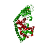
- Assembly
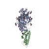
| Deposited unit | 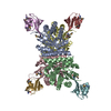
| ||||||||
|---|---|---|---|---|---|---|---|---|---|
| 1 | 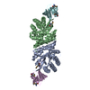
| ||||||||
| 2 | 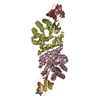
| ||||||||
| 3 |
| ||||||||
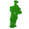
| Unit cell |
| ||||||||
| Details | The biological assembly is a tetramer in the asymmetric unit. |
-Components
| #1: Protein | Mass: 28482.160 Da / Num. of mol.: 4 Source method: isolated from a genetically manipulated source Source: (gene. exp.) Thermus thermophilus (bacteria) / Tissue: HB8 / Plasmid: pET-11A / Species (production host): Escherichia coli / Production host: Escherichia coli BL21(DE3) (bacteria) / Strain (production host): BL21(DE3) / References: UniProt: Q5SKG7#2: Protein | Mass: 7132.781 Da / Num. of mol.: 4 Source method: isolated from a genetically manipulated source Source: (gene. exp.) Thermus thermophilus (bacteria) / Tissue: HB8 / Plasmid: pET-11A / Species (production host): Escherichia coli / Production host: Escherichia coli BL21(DE3) (bacteria) / Strain (production host): BL21(DE3) / References: UniProt: Q5SKG8, UniProt: Q72KL7*PLUS#3: Water | ChemComp-HOH / | Water Mass: 18.015 Da / Num. of mol.: 359 / Source method: isolated from a natural source / Formula: H2O Mass: 18.015 Da / Num. of mol.: 359 / Source method: isolated from a natural source / Formula: H2O |
|---|
-Experimental details
-Experiment
| Experiment | Method: X-RAY DIFFRACTION / Number of used crystals: 1 |
|---|
- Sample preparation
| Crystal | Density Matthews: 2.24 Å3/Da / Density % sol: 45.05 % |
|---|---|
| Crystal grow | Temperature: 291 K / Method: microbatch / pH: 7.5 Details: PEG 4000, HEPES, pH 7.5, microbatch, temperature 291K |
-Data collection
| Diffraction | Mean temperature: 100 K |
|---|---|
| Diffraction source | Source: SYNCHROTRON / Site: SPring-8  / Beamline: BL26B1 / Wavelength: 0.97908 Å / Beamline: BL26B1 / Wavelength: 0.97908 Å |
| Detector | Type: RIGAKU RAXIS V / Detector: IMAGE PLATE / Date: Jun 29, 2005 |
| Radiation | Protocol: SINGLE WAVELENGTH / Monochromatic (M) / Laue (L): M / Scattering type: x-ray |
| Radiation wavelength | Wavelength: 0.97908 Å / Relative weight: 1 |
| Reflection | Resolution: 2.3→20 Å / Num. all: 53298 / Num. obs: 53298 / % possible obs: 98.5 % / Observed criterion σ(F): 0 / Observed criterion σ(I): 0 / Redundancy: 2.5 % / Biso Wilson estimate: 44.41 Å2 / Rmerge(I) obs: 0.066 / Rsym value: 0.059 / Net I/σ(I): 8.1 |
| Reflection shell | Resolution: 2.3→2.38 Å / Redundancy: 2.5 % / Rmerge(I) obs: 0.372 / Mean I/σ(I) obs: 2.5 / Num. unique all: 5282 / Rsym value: 0.328 / % possible all: 97.4 |
- Processing
| Software |
| |||||||||||||||||||||||||
|---|---|---|---|---|---|---|---|---|---|---|---|---|---|---|---|---|---|---|---|---|---|---|---|---|---|---|
| Refinement | Method to determine structure: MOLECULAR REPLACEMENT Starting model: PDB ENTRY 1WV2 Resolution: 2.3→19.96 Å / Isotropic thermal model: Anisotrop / Cross valid method: THROUGHOUT / σ(F): 0 / Stereochemistry target values: Engh & Huber
| |||||||||||||||||||||||||
| Displacement parameters | Biso mean: 47.9 Å2
| |||||||||||||||||||||||||
| Refine analyze |
| |||||||||||||||||||||||||
| Refinement step | Cycle: LAST / Resolution: 2.3→19.96 Å
| |||||||||||||||||||||||||
| Refine LS restraints |
| |||||||||||||||||||||||||
| LS refinement shell | Resolution: 2.3→2.38 Å / Rfactor Rfree error: 0.027
|
