 Movie
Movie Controller


+ Open data

- Basic information
| Entry | Database: PDB / ID: 2h8n | ||||||
|---|---|---|---|---|---|---|---|
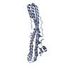
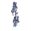
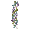
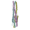
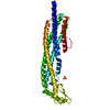
| Title | Structure of a glutamine-rich domain from histone deacetylase 4 | ||||||
 Components Components | Histone deacetylase 4 | ||||||
 Keywords Keywords | TRANSCRIPTION / alpha helix / polar zipper | ||||||
| Function / homology |  Function and homology information Function and homology informationRUNX2 regulates chondrocyte maturation / response to denervation involved in regulation of muscle adaptation / negative regulation of myotube differentiation / peptidyl-lysine deacetylation / positive regulation of protein sumoylation / negative regulation of transcription by competitive promoter binding / regulation of protein binding / protein deacetylation / cardiac muscle hypertrophy in response to stress / histone deacetylase ...RUNX2 regulates chondrocyte maturation / response to denervation involved in regulation of muscle adaptation / negative regulation of myotube differentiation / peptidyl-lysine deacetylation / positive regulation of protein sumoylation / negative regulation of transcription by competitive promoter binding / regulation of protein binding / protein deacetylation / cardiac muscle hypertrophy in response to stress / histone deacetylase / protein lysine deacetylase activity / negative regulation of glycolytic process / SUMO transferase activity / histone deacetylase activity / negative regulation of gene expression, epigenetic / B cell activation / type I interferon-mediated signaling pathway / Notch-HLH transcription pathway / potassium ion binding / protein sumoylation / RUNX3 regulates p14-ARF / histone deacetylase complex / transcription repressor complex / SUMOylation of chromatin organization proteins / response to interleukin-1 / B cell differentiation / SUMOylation of intracellular receptors / negative regulation of DNA-binding transcription factor activity / NOTCH1 Intracellular Domain Regulates Transcription / Constitutive Signaling by NOTCH1 PEST Domain Mutants / Constitutive Signaling by NOTCH1 HD+PEST Domain Mutants / histone deacetylase binding / positive regulation of DNA-binding transcription factor activity / nervous system development / DNA-binding transcription factor binding / RNA polymerase II-specific DNA-binding transcription factor binding / molecular adaptor activity / nuclear speck / chromatin remodeling / inflammatory response / RNA polymerase II cis-regulatory region sequence-specific DNA binding / positive regulation of cell population proliferation / chromatin / positive regulation of DNA-templated transcription / negative regulation of transcription by RNA polymerase II / positive regulation of transcription by RNA polymerase II / zinc ion binding / nucleoplasm / identical protein binding / nucleus / cytosol / cytoplasmSimilarity search - Function | ||||||
| Biological species |  Homo sapiens (human) Homo sapiens (human) | ||||||
| Method | X-RAY DIFFRACTION / SYNCHROTRON / SAD / Resolution: 2.6 Å | ||||||
 Authors Authors | Guo, L. / Han, A. / Bates, D.L. / Chen, L. | ||||||
 Citation Citation | Journal: Proc.Natl.Acad.Sci.Usa / Year: 2007 Title: Crystal structure of a conserved N-terminal domain of histone deacetylase 4 reveals functional insights into glutamine-rich domains. Authors: Guo, L. / Han, A. / Bates, D.L. / Cao, J. / Chen, L. | ||||||
| History |
|
- Structure visualization
| Structure viewer | Molecule: MolmilJmol/JSmol |
|---|
- Downloads & links
-Download
| PDBx/mmCIF format | 2h8n.cif.gz | 67.4 KB | Display | PDBx/mmCIF format |
|---|---|---|---|---|
| PDB format | pdb2h8n.ent.gz | 51.4 KB | Display | PDB format |
| PDBx/mmJSON format | 2h8n.json.gz | Tree view | PDBx/mmJSON format | |
| Others |  Other downloads Other downloads |
-Validation report
| Arichive directory | https://data.pdbj.org/pub/pdb/validation_reports/h8/2h8nftp://data.pdbj.org/pub/pdb/validation_reports/h8/2h8n | HTTPS FTP |
|---|
-Related structure data


-Links
 PDBj
PDBj


- Assembly
| Deposited unit | 
| ||||||||
|---|---|---|---|---|---|---|---|---|---|
| 1 |
| ||||||||
| Unit cell |
| ||||||||
| Details | The tetramer in the asymmetric unit is the biological assembly, no crystallographic symmetry is invovled in the tetramer formation. |
-Components
| #1: Protein | / HD4 Mass: 13653.348 Da / Num. of mol.: 4 / Fragment: N-terminal glutamine-rich Domain, residues 62-129 Source method: isolated from a genetically manipulated source Source: (gene. exp.) Homo sapiens (human) / Gene: HDAC4, KIAA0288 / Plasmid: pET28a / Production host:  Escherichia coli (E. coli) / Strain (production host): Rosetta PlysS / References: UniProt: P56524 Escherichia coli (E. coli) / Strain (production host): Rosetta PlysS / References: UniProt: P56524 |
|---|
-Experimental details
-Experiment
| Experiment | Method: X-RAY DIFFRACTION / Number of used crystals: 1 |
|---|
- Sample preparation
| Crystal | Density Matthews: 2.93987 Å3/Da / Density % sol: 58.161419 % Description: THE REFLECTIONS IN THE STRUCTURE FACTOR FILE WERE SCALED TO 2.3A, THE STRUCTURE WAS REFINED TO 2.6A. ALTHOUGH THE DIFFRACTION WENT TO 2.3A, DUE TO SEVERE ANISOTROPIC EFFECT, USEFUL DATA WAS ONLY TO 2.6A. |
|---|---|
| Crystal grow | Temperature: 291 K / pH: 7.5 Details: 0.825M LITHIUM SULFATE, 55mM HEPES PH 7.5, VAPOR DIFFUSION, HANGING DROP, temperature 291K, pH 7.50 |
-Data collection
| Diffraction | Mean temperature: 100 K |
|---|---|
| Diffraction source | Source: SYNCHROTRON / Site: ALS  / Beamline: 8.2.2 / Wavelength: 1.1271 / Beamline: 8.2.2 / Wavelength: 1.1271 |
| Detector | Type: ADSC QUANTUM 315 / Detector: CCD / Date: Jul 2, 2004 |
| Radiation | Protocol: SINGLE WAVELENGTH / Monochromatic (M) / Laue (L): M / Scattering type: x-ray |
| Radiation wavelength | Wavelength: 1.1271 Å / Relative weight: 1 |
| Reflection | Resolution: 2.6→6 Å / Num. obs: 19701 / % possible obs: 99 % / Observed criterion σ(I): 2 |
| Reflection shell | Resolution: 2.6→2.73 Å / % possible all: 99 |
- Processing
| Software |
| ||||||||||||||||||||||||||||||||||||||||||||||||||||||||||||||||||||||||||||||||
|---|---|---|---|---|---|---|---|---|---|---|---|---|---|---|---|---|---|---|---|---|---|---|---|---|---|---|---|---|---|---|---|---|---|---|---|---|---|---|---|---|---|---|---|---|---|---|---|---|---|---|---|---|---|---|---|---|---|---|---|---|---|---|---|---|---|---|---|---|---|---|---|---|---|---|---|---|---|---|---|---|---|
| Refinement | Method to determine structure: SAD / Resolution: 2.6→6 Å / σ(F): 2 / Stereochemistry target values: ENGH & HUBER
| ||||||||||||||||||||||||||||||||||||||||||||||||||||||||||||||||||||||||||||||||
| Displacement parameters | Biso mean: 93.34 Å2
| ||||||||||||||||||||||||||||||||||||||||||||||||||||||||||||||||||||||||||||||||
| Refinement step | Cycle: LAST / Resolution: 2.6→6 Å
| ||||||||||||||||||||||||||||||||||||||||||||||||||||||||||||||||||||||||||||||||
| Refine LS restraints |
| ||||||||||||||||||||||||||||||||||||||||||||||||||||||||||||||||||||||||||||||||
| Xplor file | Serial no: 1 / Param file: protein_rep.param |
