 Movie
Movie Controller


+ Open data

- Basic information
| Entry | Database: PDB / ID: 2h8h | ||||||
|---|---|---|---|---|---|---|---|
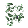
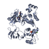
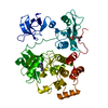
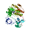
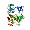
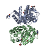
| Title | Src kinase in complex with a quinazoline inhibitor | ||||||
 Components Components | Proto-oncogene tyrosine-protein kinase Src | ||||||
 Keywords Keywords | TRANSFERASE / SRC KINASE | ||||||
| Function / homology |  Function and homology information Function and homology informationregulation of toll-like receptor 3 signaling pathway / positive regulation of non-membrane spanning protein tyrosine kinase activity / primary ovarian follicle growth / regulation of caveolin-mediated endocytosis / positive regulation of ovarian follicle development / cellular response to prolactin / positive regulation of platelet-derived growth factor receptor-beta signaling pathway / positive regulation of male germ cell proliferation / dendritic filopodium / regulation of cell projection assembly ...regulation of toll-like receptor 3 signaling pathway / positive regulation of non-membrane spanning protein tyrosine kinase activity / primary ovarian follicle growth / regulation of caveolin-mediated endocytosis / positive regulation of ovarian follicle development / cellular response to prolactin / positive regulation of platelet-derived growth factor receptor-beta signaling pathway / positive regulation of male germ cell proliferation / dendritic filopodium / regulation of cell projection assembly / regulation of cell-cell adhesion / positive regulation of dephosphorylation / response to mineralocorticoid / Regulation of commissural axon pathfinding by SLIT and ROBO / ERBB2 signaling pathway / regulation of epithelial cell migration / positive regulation of protein transport / Regulation of gap junction activity / regulation of vascular permeability / entry of bacterium into host cell / BMP receptor binding / positive regulation of lamellipodium morphogenesis / cellular response to progesterone stimulus / positive regulation of integrin activation / Activated NTRK2 signals through FYN / negative regulation of focal adhesion assembly / skeletal muscle cell proliferation / positive regulation of protein processing / Netrin mediated repulsion signals / regulation of intracellular estrogen receptor signaling pathway / intestinal epithelial cell development / CD28 co-stimulation / positive regulation of glucose metabolic process / transcytosis / Activated NTRK3 signals through PI3K / connexin binding / cellular response to fluid shear stress / focal adhesion assembly / signal complex assembly / response to acidic pH / podosome / negative regulation of telomere maintenance via telomerase / positive regulation of small GTPase mediated signal transduction / positive regulation of Ras protein signal transduction / positive regulation of podosome assembly / Regulation of RUNX1 Expression and Activity / regulation of bone resorption / branching involved in mammary gland duct morphogenesis / DCC mediated attractive signaling / negative regulation of mitochondrial depolarization / adherens junction organization / osteoclast development / myoblast proliferation / EPH-Ephrin signaling / Ephrin signaling / odontogenesis / cellular response to peptide hormone stimulus / Signal regulatory protein family interactions / cellular response to fatty acid / regulation of early endosome to late endosome transport / MET activates PTK2 signaling / oogenesis / Receptor Mediated Mitophagy / GP1b-IX-V activation signalling / Regulation of KIT signaling / Signaling by ALK / postsynaptic specialization, intracellular component / CTLA4 inhibitory signaling / Signaling by EGFR / phospholipase activator activity / leukocyte migration / DNA biosynthetic process / EPHA-mediated growth cone collapse / Fc-gamma receptor signaling pathway involved in phagocytosis / interleukin-6-mediated signaling pathway / p130Cas linkage to MAPK signaling for integrins / negative regulation of hippo signaling / positive regulation of epithelial cell migration / positive regulation of Notch signaling pathway / stress fiber assembly / negative regulation of telomerase activity / cellular response to platelet-derived growth factor stimulus / positive regulation of smooth muscle cell migration / regulation of heart rate by cardiac conduction / Recycling pathway of L1 / RUNX2 regulates osteoblast differentiation / progesterone receptor signaling pathway / dendritic growth cone / stimulatory C-type lectin receptor signaling pathway / uterus development / PECAM1 interactions / phospholipase binding / neurotrophin TRK receptor signaling pathway / Long-term potentiation / GRB2:SOS provides linkage to MAPK signaling for Integrins / RHOU GTPase cycle / platelet-derived growth factor receptor signaling pathway / FCGR activation / EPH-ephrin mediated repulsion of cells / negative regulation of anoikisSimilarity search - Function | ||||||
| Biological species |  Homo sapiens (human) Homo sapiens (human) | ||||||
| Method | X-RAY DIFFRACTION / SYNCHROTRON / MOLECULAR REPLACEMENT / Resolution: 2.2 Å | ||||||
 Authors Authors | Otterbein, L.R. / Norman, R. / Pauptit, R.A. / Rowsell, S. / Breed, J. | ||||||
 Citation Citation | Journal: J.Med.Chem. / Year: 2006 Title: N-(5-chloro-1,3-benzodioxol-4-yl)-7-[2-(4-methylpiperazin-1-yl)ethoxy]-5- (tetrahydro-2H-pyran-4-yloxy)quinazolin-4-amine, a novel, highly selective, orally available, dual-specific c-Src/Abl kinase inhibitor. Authors: Hennequin, L.F. / Allen, J. / Breed, J. / Curwen, J. / Fennell, M. / Green, T.P. / Lambert-van der Brempt, C. / Morgentin, R. / Norman, R.A. / Olivier, A. / Otterbein, L. / Ple, P.A. / Warin, N. / Costello, G. | ||||||
| History |
|
- Structure visualization
| Structure viewer | Molecule: MolmilJmol/JSmol |
|---|
- Downloads & links
-Download
| PDBx/mmCIF format | 2h8h.cif.gz | 110.8 KB | Display | PDBx/mmCIF format |
|---|---|---|---|---|
| PDB format | pdb2h8h.ent.gz | 83.4 KB | Display | PDB format |
| PDBx/mmJSON format | 2h8h.json.gz | Tree view | PDBx/mmJSON format | |
| Others |  Other downloads Other downloads |
-Validation report
| Arichive directory | https://data.pdbj.org/pub/pdb/validation_reports/h8/2h8hftp://data.pdbj.org/pub/pdb/validation_reports/h8/2h8h | HTTPS FTP |
|---|
-Related structure data
| Similar structure data |
|---|
-Links
 PDBj
PDBj


- Assembly
| Deposited unit | 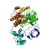
| ||||||||
|---|---|---|---|---|---|---|---|---|---|
| 1 |
| ||||||||
| Unit cell |
|
-Components
| #1: Protein | / p60-Src / c- Src / pp60c-src Mass: 59855.359 Da / Num. of mol.: 1 Source method: isolated from a genetically manipulated source Source: (gene. exp.) Homo sapiens (human) / Gene: SRC, SRC1 / Cell line (production host): SF9 cells / Production host:   Spodoptera frugiperda (fall armyworm) Spodoptera frugiperda (fall armyworm)References: UniProt: P12931, non-specific protein-tyrosine kinase |
|---|---|
| #2: Chemical | ChemComp-H8H / Saracatinib  Mass: 542.026 Da / Num. of mol.: 1 / Source method: obtained synthetically / Formula: C27H32ClN5O5 / Comment: inhibitor*YM Mass: 542.026 Da / Num. of mol.: 1 / Source method: obtained synthetically / Formula: C27H32ClN5O5 / Comment: inhibitor*YM |
| #3: Water | ChemComp-HOH / Water Mass: 18.015 Da / Num. of mol.: 221 / Source method: isolated from a natural source / Formula: H2O Mass: 18.015 Da / Num. of mol.: 221 / Source method: isolated from a natural source / Formula: H2O |
-Experimental details
-Experiment
| Experiment | Method: X-RAY DIFFRACTION / Number of used crystals: 1 |
|---|
- Sample preparation
| Crystal | Density Matthews: 2.584947 Å3/Da / Density % sol: 52.416824 % |
|---|---|
| Crystal grow | Temperature: 288 K / Method: vapor diffusion, sitting drop / pH: 6.5 Details: 50 mM PIPES pH 6.5, 10 mM DTT, 100 mM sodium chloride and 4 to 9% PEG4000 (w/v), VAPOR DIFFUSION, SITTING DROP, temperature 288K |
-Data collection
| Diffraction | Mean temperature: 100 K |
|---|---|
| Diffraction source | Source: SYNCHROTRON / Site: ESRF  / Beamline: ID14-4 / Wavelength: 1.5418 Å / Beamline: ID14-4 / Wavelength: 1.5418 Å |
| Detector | Type: ADSC QUANTUM 4 / Detector: CCD / Date: Nov 27, 2003 / Details: toroidal focusing mirror |
| Radiation | Monochromator: double crystal monochromator / Protocol: SINGLE WAVELENGTH / Monochromatic (M) / Laue (L): M / Scattering type: x-ray |
| Radiation wavelength | Wavelength: 1.5418 Å / Relative weight: 1 |
| Reflection | Resolution: 2.2→49.8 Å / Num. obs: 29365 / % possible obs: 95.49 % / Observed criterion σ(F): 1 / Observed criterion σ(I): 1 / Redundancy: 3.4 % / Rmerge(I) obs: 0.083 / Rsym value: 0.115 |
| Reflection shell | Resolution: 2.2→2.32 Å / Redundancy: 3.3 % / Rmerge(I) obs: 0.586 / Num. unique all: 4508 / Rsym value: 0.788 / % possible all: 97.2 |
- Processing
| Software |
| ||||||||||||||||||||||||||||||||||||||||||||||||||||||||||||||||||||||||||||||||||||||||||||||||||||||||||||||||||||||||||||||||||
|---|---|---|---|---|---|---|---|---|---|---|---|---|---|---|---|---|---|---|---|---|---|---|---|---|---|---|---|---|---|---|---|---|---|---|---|---|---|---|---|---|---|---|---|---|---|---|---|---|---|---|---|---|---|---|---|---|---|---|---|---|---|---|---|---|---|---|---|---|---|---|---|---|---|---|---|---|---|---|---|---|---|---|---|---|---|---|---|---|---|---|---|---|---|---|---|---|---|---|---|---|---|---|---|---|---|---|---|---|---|---|---|---|---|---|---|---|---|---|---|---|---|---|---|---|---|---|---|---|---|---|---|
| Refinement | Method to determine structure: MOLECULAR REPLACEMENT Starting model: In House Resolution: 2.2→47.67 Å / Cor.coef. Fo:Fc: 0.946 / Cor.coef. Fo:Fc free: 0.904 / SU B: 15.374 / SU ML: 0.169 / TLS residual ADP flag: LIKELY RESIDUAL / Cross valid method: THROUGHOUT / σ(F): 1 / ESU R: 0.245 / ESU R Free: 0.225 / Stereochemistry target values: MAXIMUM LIKELIHOOD / Details: HYDROGENS HAVE BEEN ADDED IN THE RIDING POSITIONS
| ||||||||||||||||||||||||||||||||||||||||||||||||||||||||||||||||||||||||||||||||||||||||||||||||||||||||||||||||||||||||||||||||||
| Solvent computation | Ion probe radii: 0.8 Å / Shrinkage radii: 0.8 Å / VDW probe radii: 1.2 Å / Solvent model: BABINET MODEL WITH MASK | ||||||||||||||||||||||||||||||||||||||||||||||||||||||||||||||||||||||||||||||||||||||||||||||||||||||||||||||||||||||||||||||||||
| Displacement parameters | Biso mean: 34.121 Å2
| ||||||||||||||||||||||||||||||||||||||||||||||||||||||||||||||||||||||||||||||||||||||||||||||||||||||||||||||||||||||||||||||||||
| Refinement step | Cycle: LAST / Resolution: 2.2→47.67 Å
| ||||||||||||||||||||||||||||||||||||||||||||||||||||||||||||||||||||||||||||||||||||||||||||||||||||||||||||||||||||||||||||||||||
| Refine LS restraints |
| ||||||||||||||||||||||||||||||||||||||||||||||||||||||||||||||||||||||||||||||||||||||||||||||||||||||||||||||||||||||||||||||||||
| LS refinement shell | Resolution: 2.2→2.257 Å / Total num. of bins used: 20
| ||||||||||||||||||||||||||||||||||||||||||||||||||||||||||||||||||||||||||||||||||||||||||||||||||||||||||||||||||||||||||||||||||
| Refinement TLS params. | Method: refined / Origin x: 19.4577 Å / Origin y: 32.8529 Å / Origin z: 66.721 Å
|
