 Movie
Movie Controller

[English] 日本語
 Yorodumi
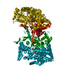
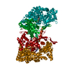
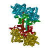
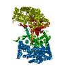
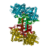
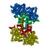
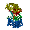
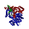
- PDB-2gm9: Structure of rabbit muscle glycogen phosphorylase in complex with... -
+ Open data

- Basic information
| Entry | Database: PDB / ID: 2gm9 | ||||||
|---|---|---|---|---|---|---|---|
| Title | Structure of rabbit muscle glycogen phosphorylase in complex with thienopyrrole | ||||||
 Components Components | Glycogen phosphorylase, muscle form | ||||||
 Keywords Keywords | TRANSFERASE / GLYCOGEN PHOSPHORYLASE | ||||||
| Function / homology |  Function and homology informationglycogen phosphorylase / glycogen phosphorylase activity / linear malto-oligosaccharide phosphorylase activity / SHG alpha-glucan phosphorylase activity / glycogen catabolic process / skeletal muscle myofibril / pyridoxal phosphate binding / nucleotide binding Function and homology informationglycogen phosphorylase / glycogen phosphorylase activity / linear malto-oligosaccharide phosphorylase activity / SHG alpha-glucan phosphorylase activity / glycogen catabolic process / skeletal muscle myofibril / pyridoxal phosphate binding / nucleotide bindingSimilarity search - Function | ||||||
| Biological species |  Oryctolagus cuniculus (rabbit) Oryctolagus cuniculus (rabbit) | ||||||
| Method | X-RAY DIFFRACTION / SYNCHROTRON / MOLECULAR REPLACEMENT / Resolution: 2.3 Å | ||||||
 Authors Authors | Otterbein, L.R. / Pannifer, A.D. / Tucker, J. / Breed, J. / Oikonomakos, N.G. / Minshull, C. / Rowsell, S. / Pauptit, R.A. | ||||||
 Citation Citation | Journal: Bioorg.Med.Chem.Lett. / Year: 2006 Title: Novel thienopyrrole glycogen phosphorylase inhibitors: synthesis, in vitro SAR and crystallographic studies. Authors: Whittamore, P.R. / Addie, M.S. / Bennett, S.N. / Birch, A.M. / Butters, M. / Godfrey, L. / Kenny, P.W. / Morley, A.D. / Murray, P.M. / Oikonomakos, N.G. / Otterbein, L.R. / Pannifer, A.D. / ...Authors: Whittamore, P.R. / Addie, M.S. / Bennett, S.N. / Birch, A.M. / Butters, M. / Godfrey, L. / Kenny, P.W. / Morley, A.D. / Murray, P.M. / Oikonomakos, N.G. / Otterbein, L.R. / Pannifer, A.D. / Parker, J.S. / Readman, K. / Siedlecki, P.S. / Schofield, P. / Stocker, A. / Taylor, M.J. / Townsend, L.A. / Whalley, D.P. / Whitehouse, J. | ||||||
| History |
|
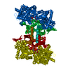
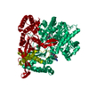
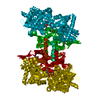
- Structure visualization
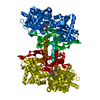
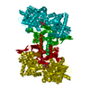
| Structure viewer | Molecule: MolmilJmol/JSmol |
|---|
- Downloads & links
-Download
| PDBx/mmCIF format | 2gm9.cif.gz | 189 KB | Display | PDBx/mmCIF format |
|---|---|---|---|---|
| PDB format | pdb2gm9.ent.gz | 145.8 KB | Display | PDB format |
| PDBx/mmJSON format | 2gm9.json.gz | Tree view | PDBx/mmJSON format | |
| Others |  Other downloads Other downloads |
-Validation report
| Arichive directory | https://data.pdbj.org/pub/pdb/validation_reports/gm/2gm9ftp://data.pdbj.org/pub/pdb/validation_reports/gm/2gm9 | HTTPS FTP |
|---|
-Related structure data


-Links
 PDBj
PDBj
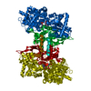
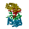
- Assembly
| Deposited unit | 
| ||||||||
|---|---|---|---|---|---|---|---|---|---|
| 1 | 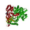
| ||||||||
| Unit cell |
| ||||||||
| Components on special symmetry positions |
| ||||||||
| Details | The second part of the biological assembly is generated by the two fold axis: |
-Components
| #1: Protein | / Myophosphorylase Mass: 95280.914 Da / Num. of mol.: 1 / Source method: isolated from a natural source / Details: Rabbit muscle / Source: (natural) Oryctolagus cuniculus (rabbit) / References: UniProt: P00489, glycogen phosphorylase |
|---|---|
| #2: Chemical | ChemComp-PLR / (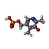  Mass: 233.158 Da / Num. of mol.: 1 / Source method: obtained synthetically / Formula: C8H12NO5P Mass: 233.158 Da / Num. of mol.: 1 / Source method: obtained synthetically / Formula: C8H12NO5P |
| #3: Chemical | ChemComp-3TH /   Mass: 345.803 Da / Num. of mol.: 1 / Source method: obtained synthetically / Formula: C16H12ClN3O2S Mass: 345.803 Da / Num. of mol.: 1 / Source method: obtained synthetically / Formula: C16H12ClN3O2S |
| #4: Water | ChemComp-HOH / Water Mass: 18.015 Da / Num. of mol.: 455 / Source method: isolated from a natural source / Formula: H2O Mass: 18.015 Da / Num. of mol.: 455 / Source method: isolated from a natural source / Formula: H2O |
-Experimental details
-Experiment
| Experiment | Method: X-RAY DIFFRACTION / Number of used crystals: 1 |
|---|
- Sample preparation
| Crystal | Density Matthews: 2.43 Å3/Da / Density % sol: 49.32 % |
|---|---|
| Crystal grow | Temperature: 289 K / Method: vapor diffusion, hanging drop / pH: 6.7 Details: 1.1mM IMP 1.1mM Spermin 10mM BES 2.9mM DTT 0.1mM EDTA, pH 6.7, VAPOR DIFFUSION, HANGING DROP, temperature 289K |
-Data collection
| Diffraction | Mean temperature: 100 K |
|---|---|
| Diffraction source | Source: SYNCHROTRON / Site: ESRF  / Beamline: ID14-4 / Wavelength: 0.93 Å / Beamline: ID14-4 / Wavelength: 0.93 Å |
| Detector | Type: ADSC QUANTUM 4 / Detector: CCD / Date: Oct 7, 2001 / Details: Toroidal Zerodur mirror |
| Radiation | Monochromator: Si111 / Protocol: SINGLE WAVELENGTH / Monochromatic (M) / Laue (L): M / Scattering type: x-ray |
| Radiation wavelength | Wavelength: 0.93 Å / Relative weight: 1 |
| Reflection | Resolution: 2.3→91.29 Å / Num. all: 40113 / Num. obs: 39447 / % possible obs: 98.34 % / Observed criterion σ(F): 0 / Observed criterion σ(I): 1 |
| Reflection shell | Resolution: 2.3→2.54 Å / % possible all: 39.7 |
- Processing
| Software |
| |||||||||||||||||||||||||||||||||||||||||||||||||||||||||||||||||||||||||||||||||||||||||||||||||||||||||||||||||||||||||||||
|---|---|---|---|---|---|---|---|---|---|---|---|---|---|---|---|---|---|---|---|---|---|---|---|---|---|---|---|---|---|---|---|---|---|---|---|---|---|---|---|---|---|---|---|---|---|---|---|---|---|---|---|---|---|---|---|---|---|---|---|---|---|---|---|---|---|---|---|---|---|---|---|---|---|---|---|---|---|---|---|---|---|---|---|---|---|---|---|---|---|---|---|---|---|---|---|---|---|---|---|---|---|---|---|---|---|---|---|---|---|---|---|---|---|---|---|---|---|---|---|---|---|---|---|---|---|---|
| Refinement | Method to determine structure: MOLECULAR REPLACEMENT Starting model: IN HOUSE MODEL Resolution: 2.3→91.29 Å / Cor.coef. Fo:Fc: 0.939 / Cor.coef. Fo:Fc free: 0.894 / SU B: 15.732 / SU ML: 0.175 / TLS residual ADP flag: LIKELY RESIDUAL / Cross valid method: THROUGHOUT / σ(F): 0 / σ(I): 1 / ESU R: 0.34 / ESU R Free: 0.244 / Stereochemistry target values: MAXIMUM LIKELIHOOD / Details: HYDROGENS HAVE BEEN ADDED IN THE RIDING POSITIONS
| |||||||||||||||||||||||||||||||||||||||||||||||||||||||||||||||||||||||||||||||||||||||||||||||||||||||||||||||||||||||||||||
| Solvent computation | Ion probe radii: 0.8 Å / Shrinkage radii: 0.8 Å / VDW probe radii: 1.2 Å / Solvent model: BABINET MODEL WITH MASK | |||||||||||||||||||||||||||||||||||||||||||||||||||||||||||||||||||||||||||||||||||||||||||||||||||||||||||||||||||||||||||||
| Displacement parameters | Biso mean: 29.819 Å2
| |||||||||||||||||||||||||||||||||||||||||||||||||||||||||||||||||||||||||||||||||||||||||||||||||||||||||||||||||||||||||||||
| Refinement step | Cycle: LAST / Resolution: 2.3→91.29 Å
| |||||||||||||||||||||||||||||||||||||||||||||||||||||||||||||||||||||||||||||||||||||||||||||||||||||||||||||||||||||||||||||
| Refine LS restraints |
| |||||||||||||||||||||||||||||||||||||||||||||||||||||||||||||||||||||||||||||||||||||||||||||||||||||||||||||||||||||||||||||
| LS refinement shell | Resolution: 2.3→2.36 Å / Total num. of bins used: 20
| |||||||||||||||||||||||||||||||||||||||||||||||||||||||||||||||||||||||||||||||||||||||||||||||||||||||||||||||||||||||||||||
| Refinement TLS params. | Method: refined / Origin x: 21.1269 Å / Origin y: 98.8218 Å / Origin z: 2.3186 Å
|
