 Movie
Movie Controller

[English] 日本語
 Yorodumi
- PDB-2ggo: Crystal Structure of glucose-1-phosphate thymidylyltransferase fr... -
+ Open data

- Basic information
| Entry | Database: PDB / ID: 2ggo | ||||||
|---|---|---|---|---|---|---|---|
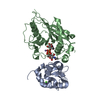
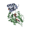
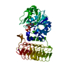
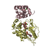
| Title | Crystal Structure of glucose-1-phosphate thymidylyltransferase from Sulfolobus tokodaii | ||||||
 Components Components | 401aa long hypothetical glucose-1-phosphate thymidylyltransferase | ||||||
 Keywords Keywords |  TRANSFERASE / BETA BARREL TRANSFERASE / BETA BARREL | ||||||
| Function / homology |  Function and homology information Function and homology informationgalactosamine-1-phosphate N-acetyltransferase / UDP-N-acetylgalactosamine diphosphorylase / UDP-N-acetylgalactosamine diphosphorylase activity / UTP-glucose-1-phosphate uridylyltransferase / UTP:glucose-1-phosphate uridylyltransferase activity / glucosamine-1-phosphate N-acetyltransferase / glucosamine-1-phosphate N-acetyltransferase activity / glucose-1-phosphate thymidylyltransferase / glucose-1-phosphate thymidylyltransferase activity / UDP-N-acetylglucosamine diphosphorylase ...galactosamine-1-phosphate N-acetyltransferase / UDP-N-acetylgalactosamine diphosphorylase / UDP-N-acetylgalactosamine diphosphorylase activity / UTP-glucose-1-phosphate uridylyltransferase / UTP:glucose-1-phosphate uridylyltransferase activity / glucosamine-1-phosphate N-acetyltransferase / glucosamine-1-phosphate N-acetyltransferase activity / glucose-1-phosphate thymidylyltransferase / glucose-1-phosphate thymidylyltransferase activity / UDP-N-acetylglucosamine diphosphorylase / UDP-N-acetylglucosamine diphosphorylase activity / UDP-N-acetylglucosamine biosynthetic process / nucleotide bindingSimilarity search - Function | ||||||
| Biological species |   Sulfolobus tokodaii (archaea) Sulfolobus tokodaii (archaea) | ||||||
| Method | X-RAY DIFFRACTION / SIRAS / Resolution: 1.8 Å | ||||||
 Authors Authors | Rajakannan, V. / Mizushima, T. / Suzuki, A. / Masui, R. / Kuramitsu, S. / Yamane, T. | ||||||
 Citation Citation | Journal: To be published Title: Crystal Structure of glucose-1-phosphate thymidylyltransferase from Sulfolobus tokodaii Authors: Rajakannan, V. / Yamane, T. | ||||||
| History |
|
- Structure visualization
| Structure viewer | Molecule: MolmilJmol/JSmol |
|---|
- Downloads & links
-Download
| PDBx/mmCIF format | 2ggo.cif.gz | 90.5 KB | Display | PDBx/mmCIF format |
|---|---|---|---|---|
| PDB format | pdb2ggo.ent.gz | 73.4 KB | Display | PDB format |
| PDBx/mmJSON format | 2ggo.json.gz | Tree view | PDBx/mmJSON format | |
| Others |  Other downloads Other downloads |
-Validation report
| Arichive directory | https://data.pdbj.org/pub/pdb/validation_reports/gg/2ggoftp://data.pdbj.org/pub/pdb/validation_reports/gg/2ggo | HTTPS FTP |
|---|
-Related structure data
| Related structure data | |
|---|---|
| Similar structure data |


-Links
 PDBj
PDBj


- Assembly
| Deposited unit | 
| |||||||||
|---|---|---|---|---|---|---|---|---|---|---|
| 1 |
| |||||||||
| 2 | 
| |||||||||
| Unit cell |
| |||||||||
| Components on special symmetry positions |
|
-Components
| #1: Protein | Mass: 44573.363 Da / Num. of mol.: 1 Source method: isolated from a genetically manipulated source Source: (gene. exp.) Sulfolobus tokodaii (archaea) / Production host:  Escherichia coli (E. coli) Escherichia coli (E. coli)References: GenBank: 15621447, UniProt: Q975F9*PLUS, glucose-1-phosphate thymidylyltransferase |
|---|---|
| #2: Water | ChemComp-HOH / Water Mass: 18.015 Da / Num. of mol.: 191 / Source method: isolated from a natural source / Formula: H2O Mass: 18.015 Da / Num. of mol.: 191 / Source method: isolated from a natural source / Formula: H2O |
-Experimental details
-Experiment
| Experiment | Method: X-RAY DIFFRACTION / Number of used crystals: 1 |
|---|
- Sample preparation
| Crystal | Density Matthews: 3.1 Å3/Da / Density % sol: 60.29 % |
|---|---|
| Crystal grow | Temperature: 293 K / Method: vapor diffusion, sitting drop / pH: 8 Details: 10% PEG 3350, 0.5M CsCl, 20mM TRIS-HCL, pH 8.0, VAPOR DIFFUSION, SITTING DROP, temperature 293K |
-Data collection
| Diffraction | Mean temperature: 100 K |
|---|---|
| Diffraction source | Source: ROTATING ANODE / Type: RIGAKU / Wavelength: 1.5418 Å |
| Detector | Type: RIGAKU RAXIS / Detector: IMAGE PLATE / Date: Dec 3, 2005 |
| Radiation | Protocol: SINGLE WAVELENGTH / Monochromatic (M) / Laue (L): M / Scattering type: x-ray |
| Radiation wavelength | Wavelength: 1.5418 Å / Relative weight: 1 |
| Reflection | Resolution: 1.8→50 Å / Num. all: 49579 / Num. obs: 48014 / % possible obs: 96.6 % / Observed criterion σ(F): 0 / Observed criterion σ(I): 0 / Redundancy: 5.3 % / Rmerge(I) obs: 0.074 |
| Reflection shell | Resolution: 1.8→1.86 Å / % possible all: 90.2 |
- Processing
| Software |
| ||||||||||||||||||||||||||||||||||||||||||||||||||||||||||||||||||||||||||||||||||||||||||
|---|---|---|---|---|---|---|---|---|---|---|---|---|---|---|---|---|---|---|---|---|---|---|---|---|---|---|---|---|---|---|---|---|---|---|---|---|---|---|---|---|---|---|---|---|---|---|---|---|---|---|---|---|---|---|---|---|---|---|---|---|---|---|---|---|---|---|---|---|---|---|---|---|---|---|---|---|---|---|---|---|---|---|---|---|---|---|---|---|---|---|---|
| Refinement | Method to determine structure: SIRAS / Resolution: 1.8→19 Å / Cor.coef. Fo:Fc: 0.955 / Cor.coef. Fo:Fc free: 0.932 / Cross valid method: THROUGHOUT / σ(F): 0 / ESU R: 0.132 / ESU R Free: 0.134 / Stereochemistry target values: MAXIMUM LIKELIHOOD / Details: HYDROGENS HAVE BEEN ADDED IN THE RIDING POSITIONS
| ||||||||||||||||||||||||||||||||||||||||||||||||||||||||||||||||||||||||||||||||||||||||||
| Solvent computation | Ion probe radii: 0.8 Å / Shrinkage radii: 0.8 Å / VDW probe radii: 1.4 Å / Solvent model: MASK | ||||||||||||||||||||||||||||||||||||||||||||||||||||||||||||||||||||||||||||||||||||||||||
| Displacement parameters | Biso mean: 44.952 Å2
| ||||||||||||||||||||||||||||||||||||||||||||||||||||||||||||||||||||||||||||||||||||||||||
| Refinement step | Cycle: LAST / Resolution: 1.8→19 Å
| ||||||||||||||||||||||||||||||||||||||||||||||||||||||||||||||||||||||||||||||||||||||||||
| Refine LS restraints |
| ||||||||||||||||||||||||||||||||||||||||||||||||||||||||||||||||||||||||||||||||||||||||||
| LS refinement shell | Resolution: 1.8→1.847 Å / Total num. of bins used: 20
|
