 Movie
Movie Controller

[English] 日本語
 Yorodumi
- PDB-2fqp: Crystal structure of a cupin domain (bp2299) from bordetella pert... -
+ Open data

- Basic information
| Entry | Database: PDB / ID: 2fqp | ||||||
|---|---|---|---|---|---|---|---|
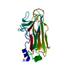
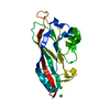
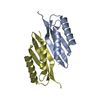
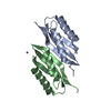
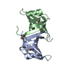
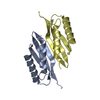
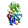
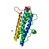
| Title | Crystal structure of a cupin domain (bp2299) from bordetella pertussis tohama i at 1.80 A resolution | ||||||
 Components Components | hypothetical protein BP2299 Hypothesis Hypothesis | ||||||
 Keywords Keywords | METAL BINDING PROTEIN / Double-stranded beta-helix fold / structural genomics / Joint Center for Structural Genomics / JCSG / Protein Structure Initiative / PSI-2 | ||||||
| Function / homology |  Function and homology information Function and homology information | ||||||
| Biological species |  Bordetella pertussis Tohama I (bacteria) Bordetella pertussis Tohama I (bacteria) | ||||||
| Method | X-RAY DIFFRACTION / SYNCHROTRON / MAD / Resolution: 1.8 Å | ||||||
 Authors Authors | Joint Center for Structural Genomics (JCSG) | ||||||
 Citation Citation | Journal: To be published Title: Crystal structure of (np_880937.1) from BORDETELLA PERTUSSIS at 1.80 A resolution Authors: Joint Center for Structural Genomics (JCSG) | ||||||
| History |
| ||||||
| Remark 999 | SEQUENCE: THE CONSTRUCT WAS EXPRESSED WITH A PURIFICATION TAG MGSDKIHHHHHHENLYFQG. THE TAG WAS ...SEQUENCE: THE CONSTRUCT WAS EXPRESSED WITH A PURIFICATION TAG MGSDKIHHHHHHENLYFQG. THE TAG WAS REMOVED WITH TEV ROTEASE LEAVING ONLY A GLYCINE (0) FOLLOWED BY THE TARGET SEQUENCE. |
- Structure visualization
| Structure viewer | Molecule: MolmilJmol/JSmol |
|---|
- Downloads & links
-Download
| PDBx/mmCIF format | 2fqp.cif.gz | 92.5 KB | Display | PDBx/mmCIF format |
|---|---|---|---|---|
| PDB format | pdb2fqp.ent.gz | 74.3 KB | Display | PDB format |
| PDBx/mmJSON format | 2fqp.json.gz | Tree view | PDBx/mmJSON format | |
| Others |  Other downloads Other downloads |
-Validation report
| Arichive directory | https://data.pdbj.org/pub/pdb/validation_reports/fq/2fqpftp://data.pdbj.org/pub/pdb/validation_reports/fq/2fqp | HTTPS FTP |
|---|
-Related structure data
| Similar structure data | |
|---|---|
| Other databases |


-Links
 PDBj
PDBj- Assembly
| Deposited unit | 
| |||||||||||||||||||||||||||||||||||||||||||||
|---|---|---|---|---|---|---|---|---|---|---|---|---|---|---|---|---|---|---|---|---|---|---|---|---|---|---|---|---|---|---|---|---|---|---|---|---|---|---|---|---|---|---|---|---|---|---|
| 1 | 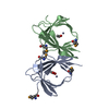
| |||||||||||||||||||||||||||||||||||||||||||||
| 2 | 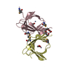
| |||||||||||||||||||||||||||||||||||||||||||||
| 3 | 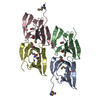
| |||||||||||||||||||||||||||||||||||||||||||||
| Unit cell |
| |||||||||||||||||||||||||||||||||||||||||||||
| Noncrystallographic symmetry (NCS) | NCS domain:
NCS domain segments: Component-ID: 1 / Ens-ID: 1 / Beg label comp-ID: ARG / Refine code: 5
|
-Components
| #1: Protein | Hypothesis Mass: 10927.838 Da / Num. of mol.: 4 Source method: isolated from a genetically manipulated source Source: (gene. exp.) Bordetella pertussis Tohama I (bacteria)Species: Bordetella pertussis / Strain: Tohama I / Gene: np_880937.1 / Plasmid: SpeedET / Production host: Escherichia coli (E. coli) / References: GenBank: 33593293, UniProt: Q7VWF8*PLUS#2: Chemical | ChemComp-ZN /   Mass: 65.409 Da / Num. of mol.: 4 / Source method: obtained synthetically / Formula: Zn Mass: 65.409 Da / Num. of mol.: 4 / Source method: obtained synthetically / Formula: Zn#3: Chemical | ChemComp-ACT / Acetate  Mass: 59.044 Da / Num. of mol.: 4 / Source method: obtained synthetically / Formula: C2H3O2 Mass: 59.044 Da / Num. of mol.: 4 / Source method: obtained synthetically / Formula: C2H3O2#4: Chemical | ChemComp-1PE / | Polyethylene glycol  Mass: 238.278 Da / Num. of mol.: 1 / Source method: obtained synthetically / Formula: C10H22O6 / Comment: precipitant*YM Mass: 238.278 Da / Num. of mol.: 1 / Source method: obtained synthetically / Formula: C10H22O6 / Comment: precipitant*YM#5: Water | ChemComp-HOH / | Water Mass: 18.015 Da / Num. of mol.: 278 / Source method: isolated from a natural source / Formula: H2O Mass: 18.015 Da / Num. of mol.: 278 / Source method: isolated from a natural source / Formula: H2O |
|---|
-Experimental details
-Experiment
| Experiment | Method: X-RAY DIFFRACTION / Number of used crystals: 1 |
|---|
- Sample preparation
| Crystal | Density Matthews: 1.83 Å3/Da / Density % sol: 36.47 % |
|---|---|
| Crystal grow | Temperature: 277 K / Method: vapor diffusion, sitting drop, nanodrop / pH: 8.5 Details: 0.2M NaOAc, 30.0% PEG-4000, 0.1M TRIS pH 8.5, VAPOR DIFFUSION,SITTING DROP,NANODROP, temperature 277K |
-Data collection
| Diffraction | Mean temperature: 100 K | |||||||||||||||||||||||||||||||||||||||||||||||||||||||||||||||||||||||||||||||||||||||||||||||||||||||||||||||||||||||||||||||||||||||||||||||||||
|---|---|---|---|---|---|---|---|---|---|---|---|---|---|---|---|---|---|---|---|---|---|---|---|---|---|---|---|---|---|---|---|---|---|---|---|---|---|---|---|---|---|---|---|---|---|---|---|---|---|---|---|---|---|---|---|---|---|---|---|---|---|---|---|---|---|---|---|---|---|---|---|---|---|---|---|---|---|---|---|---|---|---|---|---|---|---|---|---|---|---|---|---|---|---|---|---|---|---|---|---|---|---|---|---|---|---|---|---|---|---|---|---|---|---|---|---|---|---|---|---|---|---|---|---|---|---|---|---|---|---|---|---|---|---|---|---|---|---|---|---|---|---|---|---|---|---|---|---|
| Diffraction source | Source: SYNCHROTRON / Site: ALS  / Beamline: 8.3.1 / Wavelength: 1.019867, 0.979741 / Beamline: 8.3.1 / Wavelength: 1.019867, 0.979741 | |||||||||||||||||||||||||||||||||||||||||||||||||||||||||||||||||||||||||||||||||||||||||||||||||||||||||||||||||||||||||||||||||||||||||||||||||||
| Detector | Type: ADSC QUANTUM 210 / Detector: CCD / Date: Oct 11, 2005 | |||||||||||||||||||||||||||||||||||||||||||||||||||||||||||||||||||||||||||||||||||||||||||||||||||||||||||||||||||||||||||||||||||||||||||||||||||
| Radiation | Monochromator: Double Crystal Si(111) / Protocol: MAD / Monochromatic (M) / Laue (L): M / Scattering type: x-ray | |||||||||||||||||||||||||||||||||||||||||||||||||||||||||||||||||||||||||||||||||||||||||||||||||||||||||||||||||||||||||||||||||||||||||||||||||||
| Radiation wavelength |
| |||||||||||||||||||||||||||||||||||||||||||||||||||||||||||||||||||||||||||||||||||||||||||||||||||||||||||||||||||||||||||||||||||||||||||||||||||
| Reflection | Resolution: 1.8→19 Å / Num. obs: 27712 / % possible obs: 90.3 % / Redundancy: 4.1 % / Biso Wilson estimate: 13.16 Å2 / Rmerge(I) obs: 0.064 / Rsym value: 0.064 / Net I/σ(I): 11.2 | |||||||||||||||||||||||||||||||||||||||||||||||||||||||||||||||||||||||||||||||||||||||||||||||||||||||||||||||||||||||||||||||||||||||||||||||||||
| Reflection shell | Diffraction-ID: 1
|
-Phasing
| Phasing | Method: MAD |
|---|
- Processing
| Software |
| ||||||||||||||||||||||||||||||||||||||||||||||||||||||||||||||||||||||||||||||||||||||||||||||||||||||||||||||||||||||||||||||||||
|---|---|---|---|---|---|---|---|---|---|---|---|---|---|---|---|---|---|---|---|---|---|---|---|---|---|---|---|---|---|---|---|---|---|---|---|---|---|---|---|---|---|---|---|---|---|---|---|---|---|---|---|---|---|---|---|---|---|---|---|---|---|---|---|---|---|---|---|---|---|---|---|---|---|---|---|---|---|---|---|---|---|---|---|---|---|---|---|---|---|---|---|---|---|---|---|---|---|---|---|---|---|---|---|---|---|---|---|---|---|---|---|---|---|---|---|---|---|---|---|---|---|---|---|---|---|---|---|---|---|---|---|
| Refinement | Method to determine structure: MAD / Resolution: 1.8→19 Å / Cor.coef. Fo:Fc: 0.96 / Cor.coef. Fo:Fc free: 0.938 / SU B: 4.746 / SU ML: 0.079 / TLS residual ADP flag: LIKELY RESIDUAL / Cross valid method: THROUGHOUT / σ(F): 0 / ESU R: 0.147 / ESU R Free: 0.136 Stereochemistry target values: MAXIMUM LIKELIHOOD WITH PHASES Details: (1) HYDROGENS HAVE BEEN ADDED IN THE RIDING POSITIONS. (2) THERE IS A METAL ION BOUND IN EACH MONOMER. IT IS ASSIGNED AS ZN2+ BASED ON AN X-RAY EMISSION SPECTRUM AND AN X-RAY FLUORESCENCE ...Details: (1) HYDROGENS HAVE BEEN ADDED IN THE RIDING POSITIONS. (2) THERE IS A METAL ION BOUND IN EACH MONOMER. IT IS ASSIGNED AS ZN2+ BASED ON AN X-RAY EMISSION SPECTRUM AND AN X-RAY FLUORESCENCE SCAN NEAR ZN ABSORPTION EDGE. (3) THERE IS AN ACETATE ION IN EACH MONOMER BOUND NEAR ZN2+ ION. (4) THERE IS A PENTAETHYLENE GLYCOL 238 (N=5) MOLECULE BOUND IN THE STRUCTURE. THE SOURCE OF PENTAETHYLENE GLYCOL IS UNCERTAIN. IT MAY COME FROM THE CONTAMINATION OF PEG400 USED IN THE EXPERIMENT. IT IS ALSO POSSIBLE THAT WE ONLY OBSERVE THE ORDERED PART OF PEG400 AND THE REST OF PEG400 IS DISORDERED. (5) ATOM RECORD CONTAINS RESIDUAL B FACTORS ONLY
| ||||||||||||||||||||||||||||||||||||||||||||||||||||||||||||||||||||||||||||||||||||||||||||||||||||||||||||||||||||||||||||||||||
| Solvent computation | Ion probe radii: 0.8 Å / Shrinkage radii: 0.8 Å / VDW probe radii: 1.2 Å / Solvent model: BABINET MODEL WITH MASK | ||||||||||||||||||||||||||||||||||||||||||||||||||||||||||||||||||||||||||||||||||||||||||||||||||||||||||||||||||||||||||||||||||
| Displacement parameters | Biso mean: 14.089 Å2
| ||||||||||||||||||||||||||||||||||||||||||||||||||||||||||||||||||||||||||||||||||||||||||||||||||||||||||||||||||||||||||||||||||
| Refinement step | Cycle: LAST / Resolution: 1.8→19 Å
| ||||||||||||||||||||||||||||||||||||||||||||||||||||||||||||||||||||||||||||||||||||||||||||||||||||||||||||||||||||||||||||||||||
| Refine LS restraints |
| ||||||||||||||||||||||||||||||||||||||||||||||||||||||||||||||||||||||||||||||||||||||||||||||||||||||||||||||||||||||||||||||||||
| Refine LS restraints NCS | Ens-ID: 1 / Refine-ID: X-RAY DIFFRACTION
| ||||||||||||||||||||||||||||||||||||||||||||||||||||||||||||||||||||||||||||||||||||||||||||||||||||||||||||||||||||||||||||||||||
| LS refinement shell | Resolution: 1.8→1.847 Å / Total num. of bins used: 20
| ||||||||||||||||||||||||||||||||||||||||||||||||||||||||||||||||||||||||||||||||||||||||||||||||||||||||||||||||||||||||||||||||||
| Refinement TLS params. | Method: refined / Refine-ID: X-RAY DIFFRACTION
| ||||||||||||||||||||||||||||||||||||||||||||||||||||||||||||||||||||||||||||||||||||||||||||||||||||||||||||||||||||||||||||||||||
| Refinement TLS group | Refine-ID: X-RAY DIFFRACTION / Selection: ALL
|
