 Movie
Movie Controller

[English] 日本語
 Yorodumi
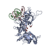
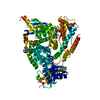
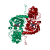
- PDB-2fo1: Crystal Structure of the CSL-Notch-Mastermind ternary complex bou... -
+ Open data

- Basic information
| Entry | Database: PDB / ID: 2fo1 | ||||||
|---|---|---|---|---|---|---|---|
| Title | Crystal Structure of the CSL-Notch-Mastermind ternary complex bound to DNA | ||||||
 Components Components |
| ||||||
 Keywords Keywords | GENE REGULATION/SIGNALLING PROTEIN/DNA /  beta-barrel / PROTEIN-DNA COMPLEX / DOUBLE HELIX / ankyrin repeat / GENE REGULATION-SIGNALLING PROTEIN-DNA COMPLEX beta-barrel / PROTEIN-DNA COMPLEX / DOUBLE HELIX / ankyrin repeat / GENE REGULATION-SIGNALLING PROTEIN-DNA COMPLEX | ||||||
| Function / homology |  Function and homology information Function and homology informationstem cell fate determination / Pre-NOTCH Processing in Golgi / NOTCH4 Activation and Transmission of Signal to the Nucleus / Negative regulation of NOTCH4 signaling / regulation of mesodermal cell fate specification / dauer exit / positive regulation of vulval development / CSL-Notch-Mastermind transcription factor complex / cell projection morphogenesis / positive regulation of mesodermal cell fate specification ...stem cell fate determination / Pre-NOTCH Processing in Golgi / NOTCH4 Activation and Transmission of Signal to the Nucleus / Negative regulation of NOTCH4 signaling / regulation of mesodermal cell fate specification / dauer exit / positive regulation of vulval development / CSL-Notch-Mastermind transcription factor complex / cell projection morphogenesis / positive regulation of mesodermal cell fate specification / vulval development / regulation of vulval development / oocyte growth / nematode larval development / germ-line stem cell division / regulation of basement membrane organization / egg-laying behavior / regulation of cell fate specification / cell fate determination / sleep / Notch binding / cell fate specification / positive regulation of stem cell proliferation / Notch signaling pathway / transcription coactivator binding / positive regulation of miRNA transcription / RNA polymerase II transcription regulator complex / transmembrane signaling receptor activity / double-stranded DNA binding / regulation of gene expression / DNA-binding transcription activator activity, RNA polymerase II-specific / DNA-binding transcription factor binding / RNA polymerase II-specific DNA-binding transcription factor binding / sequence-specific DNA binding / transcription coactivator activity / DNA-binding transcription factor activity, RNA polymerase II-specific / apical plasma membrane / RNA polymerase II cis-regulatory region sequence-specific DNA binding / calcium ion binding / chromatin / regulation of transcription by RNA polymerase II / positive regulation of DNA-templated transcription / negative regulation of transcription by RNA polymerase II / positive regulation of transcription by RNA polymerase II / nucleus / plasma membraneSimilarity search - Function | ||||||
| Biological species |  Caenorhabditis elegans (invertebrata) Caenorhabditis elegans (invertebrata) | ||||||
| Method | X-RAY DIFFRACTION / SYNCHROTRON / SIRAS, MIRAS, molecular replacement / Resolution: 3.12 Å | ||||||
 Authors Authors | Wilson, J.J. / Kovall, R.A. | ||||||
 Citation Citation | Journal: Cell(Cambridge,Mass.) / Year: 2006 Title: Crystal structure of the CSL-Notch-Mastermind ternary complex bound to DNA. Authors: Wilson, J.J. / Kovall, R.A. | ||||||
| History |
|
- Structure visualization
| Structure viewer | Molecule: MolmilJmol/JSmol |
|---|
- Downloads & links
-Download
| PDBx/mmCIF format | 2fo1.cif.gz | 181.2 KB | Display | PDBx/mmCIF format |
|---|---|---|---|---|
| PDB format | pdb2fo1.ent.gz | 136.2 KB | Display | PDB format |
| PDBx/mmJSON format | 2fo1.json.gz | Tree view | PDBx/mmJSON format | |
| Others |  Other downloads Other downloads |
-Validation report
| Arichive directory | https://data.pdbj.org/pub/pdb/validation_reports/fo/2fo1ftp://data.pdbj.org/pub/pdb/validation_reports/fo/2fo1 | HTTPS FTP |
|---|
-Related structure data


-Links
 PDBj
PDBj


- Assembly
| Deposited unit | 
| ||||||||
|---|---|---|---|---|---|---|---|---|---|
| 1 |
| ||||||||
| Unit cell |
|
-Components
| #1: DNA chain | Mass: 4673.059 Da / Num. of mol.: 1 / Source method: obtained synthetically Details: THE SEQUENCE COMES FROM A REGION WITHIN THE MAMMALIAN HES-1 PROMOTER |
|---|---|
| #2: DNA chain | Mass: 4503.949 Da / Num. of mol.: 1 / Source method: obtained synthetically Details: THE SEQUENCE COMES FROM A REGION WITHIN THE MAMMALIAN HES-1 PROMOTER |
| #3: Protein | Mass: 53761.641 Da / Num. of mol.: 1 / Fragment: CORE (RESIDUES 192-663) Source method: isolated from a genetically manipulated source Source: (gene. exp.) Caenorhabditis elegans (invertebrata) / Gene: LAG-1 / Plasmid: PGEX6P-1 / Production host:  Escherichia coli (E. coli) / Strain (production host): ROSETTA2(DE3)PLYSS / References: GenBank: 22532887, UniProt: V6CLJ5*PLUS Escherichia coli (E. coli) / Strain (production host): ROSETTA2(DE3)PLYSS / References: GenBank: 22532887, UniProt: V6CLJ5*PLUS |
| #4: Protein | Mass: 9755.596 Da / Num. of mol.: 1 / Fragment: CONSERVED N-TERMINUS (49-132) Source method: isolated from a genetically manipulated source Source: (gene. exp.) Caenorhabditis elegans (invertebrata) / Gene: sel-8, lag-3 / Plasmid: pET28A pSMT variant / Production host: Escherichia coli (E. coli) / Strain (production host): ROSETTA2(DE3)PLYSS / References: UniProt: Q09260 |
| #5: Protein | Mass: 42457.406 Da / Num. of mol.: 1 / Fragment: RAM AND ANK REPEAT DOMAINS (931-1303) Source method: isolated from a genetically manipulated source Source: (gene. exp.) Caenorhabditis elegans (invertebrata) / Gene: lin-12 / Plasmid: pGEX4T-2 / Production host: Escherichia coli (E. coli) / Strain (production host): ROSETTA2(DE3)PLYSS / References: UniProt: P14585 |
-Experimental details
-Experiment
| Experiment | Method: X-RAY DIFFRACTION / Number of used crystals: 1 |
|---|
- Sample preparation
| Crystal | Density Matthews: 3.39 Å3/Da / Density % sol: 63.68 % | ||||||||||||||||||||||||||||||||
|---|---|---|---|---|---|---|---|---|---|---|---|---|---|---|---|---|---|---|---|---|---|---|---|---|---|---|---|---|---|---|---|---|---|
| Crystal grow | Temperature: 277 K / Method: microbatch / pH: 5.5 Details: 10% PEG 10K, 0.15M Ammonium Acetate, 0.1M BisTris, 10% ethylene glycol, pH 5.5, MICROBATCH, temperature 277K | ||||||||||||||||||||||||||||||||
| Components of the solutions |
|
-Data collection
| Diffraction | Mean temperature: 200 K |
|---|---|
| Diffraction source | Source: SYNCHROTRON / Site: APS  / Beamline: 17-ID / Wavelength: 0.9796 Å / Beamline: 17-ID / Wavelength: 0.9796 Å |
| Detector | Type: MAR CCD 165 mm / Detector: CCD / Date: Jul 1, 2005 |
| Radiation | Monochromator: Si(111) double-crystal system / Protocol: SINGLE WAVELENGTH / Monochromatic (M) / Laue (L): M / Scattering type: x-ray |
| Radiation wavelength | Wavelength: 0.9796 Å / Relative weight: 1 |
| Reflection | Resolution: 3.12→50 Å / Num. all: 52029 / Num. obs: 47404 / % possible obs: 98.3 % / Observed criterion σ(F): 1 / Observed criterion σ(I): 3 / Redundancy: 13.5 % / Rsym value: 0.086 / Net I/σ(I): 25 |
| Reflection shell | Resolution: 3.12→3.23 Å / Redundancy: 9.1 % / Mean I/σ(I) obs: 3.2 / Rsym value: 0.465 / % possible all: 88.2 |
- Processing
| Software |
| ||||||||||||||||||||||||||||||||||||||||||||||||||||||||||||||||||||||||||||||||
|---|---|---|---|---|---|---|---|---|---|---|---|---|---|---|---|---|---|---|---|---|---|---|---|---|---|---|---|---|---|---|---|---|---|---|---|---|---|---|---|---|---|---|---|---|---|---|---|---|---|---|---|---|---|---|---|---|---|---|---|---|---|---|---|---|---|---|---|---|---|---|---|---|---|---|---|---|---|---|---|---|---|
| Refinement | Method to determine structure: SIRAS, MIRAS, molecular replacement Starting model: PDB ENTRY 1TTU PDB ENTRY 1OT8 Resolution: 3.12→43.51 Å / Rfactor Rfree error: 0.005 / Data cutoff high absF: 68268.33 / Data cutoff high rms absF: 68268.33 / Data cutoff low absF: 0 / Isotropic thermal model: RESTRAINED / Cross valid method: THROUGHOUT / σ(F): 0 / Stereochemistry target values: Engh & Huber
| ||||||||||||||||||||||||||||||||||||||||||||||||||||||||||||||||||||||||||||||||
| Solvent computation | Solvent model: FLAT MODEL / Bsol: 41.6717 Å2 / ksol: 0.201211 e/Å3 | ||||||||||||||||||||||||||||||||||||||||||||||||||||||||||||||||||||||||||||||||
| Displacement parameters | Biso mean: 103.8 Å2
| ||||||||||||||||||||||||||||||||||||||||||||||||||||||||||||||||||||||||||||||||
| Refine analyze |
| ||||||||||||||||||||||||||||||||||||||||||||||||||||||||||||||||||||||||||||||||
| Refinement step | Cycle: LAST / Resolution: 3.12→43.51 Å
| ||||||||||||||||||||||||||||||||||||||||||||||||||||||||||||||||||||||||||||||||
| Refine LS restraints |
| ||||||||||||||||||||||||||||||||||||||||||||||||||||||||||||||||||||||||||||||||
| LS refinement shell | Resolution: 3.12→3.32 Å / Rfactor Rfree error: 0.02 / Total num. of bins used: 6
| ||||||||||||||||||||||||||||||||||||||||||||||||||||||||||||||||||||||||||||||||
| Xplor file |
|
