Resolution of AP sites via the single-nucleotide replacement pathway / immunoglobulin heavy chain V-D-J recombination / Resolution of AP sites via the multiple-nucleotide patch replacement pathway / Abasic sugar-phosphate removal via the single-nucleotide replacement pathway / APEX1-Independent Resolution of AP Sites via the Single Nucleotide Replacement Pathway / Lyases; Carbon-oxygen lyases; Other carbon-oxygen lyases / homeostasis of number of cells / PCNA-Dependent Long Patch Base Excision Repair / 5'-deoxyribose-5-phosphate lyase activity / POLB-Dependent Long Patch Base Excision Repair ...Resolution of AP sites via the single-nucleotide replacement pathway / immunoglobulin heavy chain V-D-J recombination / Resolution of AP sites via the multiple-nucleotide patch replacement pathway / Abasic sugar-phosphate removal via the single-nucleotide replacement pathway / APEX1-Independent Resolution of AP Sites via the Single Nucleotide Replacement Pathway / Lyases; Carbon-oxygen lyases; Other carbon-oxygen lyases / homeostasis of number of cells / PCNA-Dependent Long Patch Base Excision Repair / 5'-deoxyribose-5-phosphate lyase activity / POLB-Dependent Long Patch Base Excision Repair / pyrimidine dimer repair / response to hyperoxia / somatic hypermutation of immunoglobulin genes / lymph node development / salivary gland morphogenesis / class I DNA-(apurinic or apyrimidinic site) endonuclease activity / base-excision repair, gap-filling / spleen development / DNA-(apurinic or apyrimidinic site) endonuclease activity / DNA-(apurinic or apyrimidinic site) lyase / spindle microtubule / response to gamma radiation / base-excision repair / DNA-templated DNA replication / double-strand break repair via nonhomologous end joining / intrinsic apoptotic signaling pathway in response to DNA damage / microtubule binding / neuron apoptotic process / response to ethanol / in utero embryonic development / microtubule / damaged DNA binding / DNA-directed DNA polymerase / DNA-directed DNA polymerase activity / lyase activity / Ub-specific processing proteases / inflammatory response / DNA repair / DNA damage response / enzyme binding / protein-containing complex / nucleoplasm / metal ion binding / nucleus / cytoplasm Similarity search - Function
Beta Polymerase; domain 3 / DNA polymerase, thumb domain / DNA polymerase family X, beta-like / DNA polymerase beta, N-terminal domain-like / DNA polymerase beta, palm domain / DNA polymerase beta palm / DNA polymerase lambda, fingers domain / Fingers domain of DNA polymerase lambda / DNA-directed DNA polymerase X / DNA polymerase beta-like, N-terminal domain ...Beta Polymerase; domain 3 / DNA polymerase, thumb domain / DNA polymerase family X, beta-like / DNA polymerase beta, N-terminal domain-like / DNA polymerase beta, palm domain / DNA polymerase beta palm / DNA polymerase lambda, fingers domain / Fingers domain of DNA polymerase lambda / DNA-directed DNA polymerase X / DNA polymerase beta-like, N-terminal domain / Helix-hairpin-helix domain / DNA polymerase X family / DNA polymerase lambda lyase domain superfamily / DNA polymerase family X, binding site / DNA polymerase family X signature. / DNA polymerase family X / DNA polymerase beta, thumb domain / DNA polymerase beta thumb / DNA polymerase, thumb domain superfamily / Beta Polymerase, domain 2 / Helix-hairpin-helix DNA-binding motif, class 1 / Helix-hairpin-helix DNA-binding motif class 1 / Beta Polymerase; domain 2 / Nucleotidyltransferase superfamily / 5' to 3' exonuclease, C-terminal subdomain / DNA polymerase; domain 1 / 2-Layer Sandwich / Orthogonal Bundle / Mainly Alpha / Alpha Beta Similarity search - Domain/homology
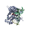
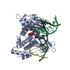
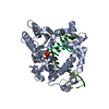
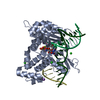
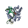
2'-DEOXYURIDINE 5'-ALPHA,BETA-IMIDO-TRIPHOSPHATE / DNA / DNA (> 10) / DNA polymerase beta Similarity search - Component
Biological species
Homo sapiens (human)
Method
X-RAY DIFFRACTION / MOLECULAR REPLACEMENT / Resolution: 2.2 Å
Mass: 4853.158 Da / Num. of mol.: 1 / Source method: obtained synthetically
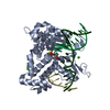
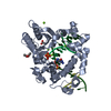
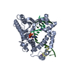
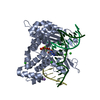
#2: DNA chain
5'-D(*GP*CP*TP*GP*AP*TP*GP*CP*GP*C)-3'
Mass: 3061.004 Da / Num. of mol.: 1 / Source method: obtained synthetically
#3: DNA chain
5'-D(P*GP*TP*CP*GP*G)-3'
Mass: 1536.035 Da / Num. of mol.: 1 / Source method: obtained synthetically
-
Protein , 1 types, 1 molecules A
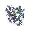
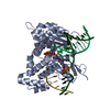
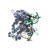
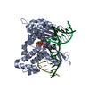
#4: Protein
DNAPolymeraseBeta /
Mass: 38241.672 Da / Num. of mol.: 1 Source method: isolated from a genetically manipulated source Source: (gene. exp.) Homo sapiens (human) / Gene: POLB / Production host: Escherichia coli (E. coli) / References: UniProt: P06746, DNA-directed DNA polymerase
In the structure databanks used in Yorodumi, some data are registered as the other names, "COVID-19 virus" and "2019-nCoV". Here are the details of the virus and the list of structure data.
Jan 31, 2019. EMDB accession codes are about to change! (news from PDBe EMDB page)
EMDB accession codes are about to change! (news from PDBe EMDB page)
The allocation of 4 digits for EMDB accession codes will soon come to an end. Whilst these codes will remain in use, new EMDB accession codes will include an additional digit and will expand incrementally as the available range of codes is exhausted. The current 4-digit format prefixed with “EMD-” (i.e. EMD-XXXX) will advance to a 5-digit format (i.e. EMD-XXXXX), and so on. It is currently estimated that the 4-digit codes will be depleted around Spring 2019, at which point the 5-digit format will come into force.
The EM Navigator/Yorodumi systems omit the EMD- prefix.
Related info.:Q: What is EMD? / ID/Accession-code notation in Yorodumi/EM Navigator
Yorodumi is a browser for structure data from EMDB, PDB, SASBDB, etc.
This page is also the successor to EM Navigator detail page, and also detail information page/front-end page for Omokage search.
The word "yorodu" (or yorozu) is an old Japanese word meaning "ten thousand". "mi" (miru) is to see.
Related info.:EMDB / PDB / SASBDB / Comparison of 3 databanks / Yorodumi Search / Aug 31, 2016. New EM Navigator & Yorodumi / Yorodumi Papers / Jmol/JSmol / Function and homology information / Changes in new EM Navigator and Yorodumi
 Movie
Movie Controller


 Open data
 Basic information
 Components
 Keywords
 Nucleotidyl Transferase / TRANSFERASE-DNA COMPLEX
 Function and homology information
 Authors
 Citation
 Structure visualization
 Downloads & links
 Other downloads
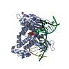


 PDBj
PDBj


 Assembly
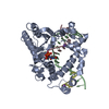


 Mass: 24.305 Da / Num. of mol.: 1 / Source method: obtained synthetically / Formula: Mg
 Mass: 22.990 Da / Num. of mol.: 3 / Source method: obtained synthetically / Formula: Na
 Mass: 467.157 Da / Num. of mol.: 1 / Source method: obtained synthetically / Formula: C9H16N3O13P3
 Sample preparation
 Processing