 Movie
Movie Controller

[English] 日本語
 Yorodumi
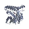
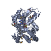
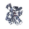
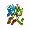
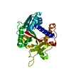
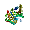
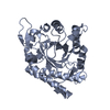
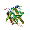
- PDB-2fk8: Crystal structure of Hma (MmaA4) from Mycobacterium tuberculosis ... -
+ Open data

- Basic information
| Entry | Database: PDB / ID: 2fk8 | ||||||
|---|---|---|---|---|---|---|---|
| Title | Crystal structure of Hma (MmaA4) from Mycobacterium tuberculosis complexed with S-adenosylmethionine | ||||||
 Components Components | methoxy mycolic acid synthase 4 | ||||||
 Keywords Keywords |  TRANSFERASE / S-ADENOSYLMETHIONINE-DEPENDENT METHYLTRANSFERASE FOLD TRANSFERASE / S-ADENOSYLMETHIONINE-DEPENDENT METHYLTRANSFERASE FOLD | ||||||
| Function / homology |  Function and homology information Function and homology informationmycolic acid biosynthetic process / S-adenosylmethionine-dependent methyltransferase activity / Transferases; Transferring one-carbon groups; Methyltransferases / peptidoglycan-based cell wall / methyltransferase activity / methylation / plasma membraneSimilarity search - Function | ||||||
| Biological species |   Mycobacterium tuberculosis (bacteria) Mycobacterium tuberculosis (bacteria) | ||||||
| Method | X-RAY DIFFRACTION / SYNCHROTRON / MOLECULAR REPLACEMENT / Resolution: 2 Å | ||||||
 Authors Authors | Boissier, F. / Guillet, V. / Mourey, L. | ||||||
 Citation Citation | Journal: J.Biol.Chem. / Year: 2006 Title: Further insight into S-adenosylmethionine-dependent methyltransferases: structural characterization of Hma, an enzyme essential for the biosynthesis of oxygenated mycolic acids in Mycobacterium tuberculosis. Authors: Boissier, F. / Bardou, F. / Guillet, V. / Uttenweiler-Joseph, S. / Daffe, M. / Quemard, A. / Mourey, L. | ||||||
| History |
|
- Structure visualization
| Structure viewer | Molecule: MolmilJmol/JSmol |
|---|
- Downloads & links
-Download
| PDBx/mmCIF format | 2fk8.cif.gz | 75.9 KB | Display | PDBx/mmCIF format |
|---|---|---|---|---|
| PDB format | pdb2fk8.ent.gz | 54.2 KB | Display | PDB format |
| PDBx/mmJSON format | 2fk8.json.gz | Tree view | PDBx/mmJSON format | |
| Others |  Other downloads Other downloads |
-Validation report
| Arichive directory | https://data.pdbj.org/pub/pdb/validation_reports/fk/2fk8ftp://data.pdbj.org/pub/pdb/validation_reports/fk/2fk8 | HTTPS FTP |
|---|
-Related structure data
| Related structure data |  2fk7SC S: Starting model for refinement C: citing same article ( |
|---|---|
| Similar structure data |


-Links
 PDBj
PDBj
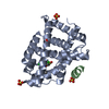
- Assembly
| Deposited unit | 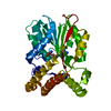
| ||||||||
|---|---|---|---|---|---|---|---|---|---|
| 1 |
| ||||||||
| Unit cell |
| ||||||||
| Details | The biological assembly is the monomer found in the asymmetric unit |
-Components
| #1: Protein | Mass: 36521.305 Da / Num. of mol.: 1 Source method: isolated from a genetically manipulated source Source: (gene. exp.) Mycobacterium tuberculosis (bacteria) / Strain: H37Rv / Gene: mmaA4 (Rv0642c) / Plasmid: pET-15b / Production host: Escherichia coli (E. coli) / Strain (production host): BL21(DE3)pLysSReferences: GenBank: 13880191, UniProt: Q79FX8*PLUS, Transferases; Transferring one-carbon groups; Methyltransferases |
|---|---|
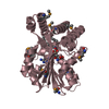
| #2: Chemical | ChemComp-SAM / S-Adenosyl methionine  Mass: 398.437 Da / Num. of mol.: 1 / Source method: obtained synthetically / Formula: C15H22N6O5S Mass: 398.437 Da / Num. of mol.: 1 / Source method: obtained synthetically / Formula: C15H22N6O5S |
| #3: Water | ChemComp-HOH / Water Mass: 18.015 Da / Num. of mol.: 153 / Source method: isolated from a natural source / Formula: H2O Mass: 18.015 Da / Num. of mol.: 153 / Source method: isolated from a natural source / Formula: H2O |
-Experimental details
-Experiment
| Experiment | Method: X-RAY DIFFRACTION / Number of used crystals: 1 |
|---|
- Sample preparation
| Crystal | Density Matthews: 2.67 Å3/Da / Density % sol: 53.88 % |
|---|---|
| Crystal grow | Temperature: 285 K / Method: vapor diffusion, hanging drop / pH: 5 Details: 4-28% PEG 3350, pH 5.0, VAPOR DIFFUSION, HANGING DROP, temperature 285.0K |
-Data collection
| Diffraction | Mean temperature: 100 K |
|---|---|
| Diffraction source | Source: SYNCHROTRON / Site: ESRF  / Beamline: ID14-4 / Wavelength: 0.979171 Å / Beamline: ID14-4 / Wavelength: 0.979171 Å |
| Detector | Type: ADSC QUANTUM 4 / Detector: CCD / Date: Dec 6, 2004 / Details: Toroidal mirror |
| Radiation | Monochromator: Double crystal, Si(111) or Si(311) / Protocol: SINGLE WAVELENGTH / Monochromatic (M) / Laue (L): M / Scattering type: x-ray |
| Radiation wavelength | Wavelength: 0.979171 Å / Relative weight: 1 |
| Reflection | Resolution: 2→30 Å / Num. all: 25549 / Num. obs: 25549 / % possible obs: 97.7 % / Redundancy: 3.9 % / Biso Wilson estimate: 30.6 Å2 / Rsym value: 0.085 / Net I/σ(I): 4.8 |
| Reflection shell | Resolution: 2→2.11 Å / Redundancy: 3.2 % / Mean I/σ(I) obs: 2.9 / Num. unique all: 3284 / Rsym value: 0.223 / % possible all: 88.1 |
- Processing
| Software |
| ||||||||||||||||||||||||||||||||||||||||||||||||||||||||||||||||||||||||||||||||||||||||||||||||||||||||||||||||||||||||||||||||||||||||||||||||||||||||||||||||||||||||||
|---|---|---|---|---|---|---|---|---|---|---|---|---|---|---|---|---|---|---|---|---|---|---|---|---|---|---|---|---|---|---|---|---|---|---|---|---|---|---|---|---|---|---|---|---|---|---|---|---|---|---|---|---|---|---|---|---|---|---|---|---|---|---|---|---|---|---|---|---|---|---|---|---|---|---|---|---|---|---|---|---|---|---|---|---|---|---|---|---|---|---|---|---|---|---|---|---|---|---|---|---|---|---|---|---|---|---|---|---|---|---|---|---|---|---|---|---|---|---|---|---|---|---|---|---|---|---|---|---|---|---|---|---|---|---|---|---|---|---|---|---|---|---|---|---|---|---|---|---|---|---|---|---|---|---|---|---|---|---|---|---|---|---|---|---|---|---|---|---|---|---|---|
| Refinement | Method to determine structure: MOLECULAR REPLACEMENT Starting model: pdb entry 2FK7 Resolution: 2→30 Å / Cor.coef. Fo:Fc: 0.957 / Cor.coef. Fo:Fc free: 0.921 / SU B: 6.911 / SU ML: 0.099 / TLS residual ADP flag: LIKELY RESIDUAL Isotropic thermal model: isotropic + anisotropic in the last stages of refinement (TLS parameters were refined using a single group) Cross valid method: THROUGHOUT / ESU R: 0.15 / ESU R Free: 0.155 / Stereochemistry target values: MAXIMUM LIKELIHOOD / Details: HYDROGENS HAVE BEEN ADDED IN THE RIDING POSITIONS
| ||||||||||||||||||||||||||||||||||||||||||||||||||||||||||||||||||||||||||||||||||||||||||||||||||||||||||||||||||||||||||||||||||||||||||||||||||||||||||||||||||||||||||
| Solvent computation | Ion probe radii: 0.8 Å / Shrinkage radii: 0.8 Å / VDW probe radii: 1.2 Å / Solvent model: BABINET MODEL WITH MASK | ||||||||||||||||||||||||||||||||||||||||||||||||||||||||||||||||||||||||||||||||||||||||||||||||||||||||||||||||||||||||||||||||||||||||||||||||||||||||||||||||||||||||||
| Displacement parameters | Biso mean: 36.975 Å2
| ||||||||||||||||||||||||||||||||||||||||||||||||||||||||||||||||||||||||||||||||||||||||||||||||||||||||||||||||||||||||||||||||||||||||||||||||||||||||||||||||||||||||||
| Refinement step | Cycle: LAST / Resolution: 2→30 Å
| ||||||||||||||||||||||||||||||||||||||||||||||||||||||||||||||||||||||||||||||||||||||||||||||||||||||||||||||||||||||||||||||||||||||||||||||||||||||||||||||||||||||||||
| Refine LS restraints |
| ||||||||||||||||||||||||||||||||||||||||||||||||||||||||||||||||||||||||||||||||||||||||||||||||||||||||||||||||||||||||||||||||||||||||||||||||||||||||||||||||||||||||||
| LS refinement shell | Resolution: 2→2.052 Å / Total num. of bins used: 20
| ||||||||||||||||||||||||||||||||||||||||||||||||||||||||||||||||||||||||||||||||||||||||||||||||||||||||||||||||||||||||||||||||||||||||||||||||||||||||||||||||||||||||||
| Refinement TLS params. | Method: refined / Origin x: 24.1462 Å / Origin y: -4.9533 Å / Origin z: 16.7397 Å
|
