 Movie
Movie Controller


+ Open data

- Basic information
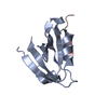
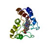
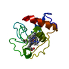
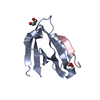
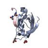
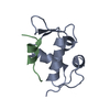
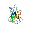
| Entry | Database: PDB / ID: 2fe5 | ||||||
|---|---|---|---|---|---|---|---|
| Title | The Crystal Structure of the Second PDZ Domain of Human DLG3 | ||||||
 Components Components | Presynaptic protein SAP102 Synapse Synapse | ||||||
 Keywords Keywords | STRUCTURAL PROTEIN / PDZ domain / DLG3 / Human / Structural Genomics / Structural Genomics Consortium / SGC | ||||||
| Function / homology |  Function and homology information Function and homology informationestablishment of planar polarity / NrCAM interactions / receptor localization to synapse / Synaptic adhesion-like molecules / establishment or maintenance of epithelial cell apical/basal polarity / Assembly and cell surface presentation of NMDA receptors / Neurexins and neuroligins / Activation of Ca-permeable Kainate Receptor / receptor clustering / Negative regulation of NMDA receptor-mediated neuronal transmission ...establishment of planar polarity / NrCAM interactions / receptor localization to synapse / Synaptic adhesion-like molecules / establishment or maintenance of epithelial cell apical/basal polarity / Assembly and cell surface presentation of NMDA receptors / Neurexins and neuroligins / Activation of Ca-permeable Kainate Receptor / receptor clustering / Negative regulation of NMDA receptor-mediated neuronal transmission / Unblocking of NMDA receptors, glutamate binding and activation / regulation of postsynaptic membrane neurotransmitter receptor levels / AMPA glutamate receptor complex / Long-term potentiation / bicellular tight junction / phosphatase binding / positive regulation of protein tyrosine kinase activity / Ras activation upon Ca2+ influx through NMDA receptor / postsynaptic density membrane / adherens junction / neuromuscular junction / cell-cell adhesion / kinase binding / chemical synaptic transmission / RAF/MAP kinase cascade / basolateral plasma membrane / neuron projection / negative regulation of cell population proliferation / glutamatergic synapse / ubiquitin protein ligase binding / extracellular space / plasma membrane / cytosolSimilarity search - Function | ||||||
| Biological species |  Homo sapiens (human) Homo sapiens (human) | ||||||
| Method | X-RAY DIFFRACTION / SYNCHROTRON / MOLECULAR REPLACEMENT / Resolution: 1.1 Å | ||||||
 Authors Authors | Ugochukwu, E. / Phillips, C. / Schoch, G. / Berridge, G. / Salah, E. / Colebrook, S. / Smee, C. / Savitsky, P. / Bray, J. / Elkins, J. ...Ugochukwu, E. / Phillips, C. / Schoch, G. / Berridge, G. / Salah, E. / Colebrook, S. / Smee, C. / Savitsky, P. / Bray, J. / Elkins, J. / Doyle, D. / Bunkoczi, G. / Debreczeni, J. / Turnbull, A. / Gorrec, F. / von Delft, F. / Sundstrom, M. / Arrowsmith, C. / Weigelt, J. / Edwards, A. / Structural Genomics Consortium (SGC) | ||||||
 Citation Citation | Journal: To be Published Title: The Crystal Structure of the Second PDZ Domain of Human DLG3 Authors: Ugochukwu, E. / Phillips, C. / Schoch, G. / Berridge, G. / Salah, E. / Colebrook, S. / Smee, C. / Savitsky, P. / Bray, J. / Elkins, J. / Doyle, D. / Bunkoczi, G. / Debreczeni, J. / Turnbull, ...Authors: Ugochukwu, E. / Phillips, C. / Schoch, G. / Berridge, G. / Salah, E. / Colebrook, S. / Smee, C. / Savitsky, P. / Bray, J. / Elkins, J. / Doyle, D. / Bunkoczi, G. / Debreczeni, J. / Turnbull, A. / Gorrec, F. / von Delft, F. / Sundstrom, M. / Arrowsmith, C. / Weigelt, J. / Edwards, A. / Structural Genomics Consortium (SGC) | ||||||
| History |
|
- Structure visualization
| Structure viewer | Molecule: MolmilJmol/JSmol |
|---|
- Downloads & links
-Download
| PDBx/mmCIF format | 2fe5.cif.gz | 58.7 KB | Display | PDBx/mmCIF format |
|---|---|---|---|---|
| PDB format | pdb2fe5.ent.gz | 42.8 KB | Display | PDB format |
| PDBx/mmJSON format | 2fe5.json.gz | Tree view | PDBx/mmJSON format | |
| Others |  Other downloads Other downloads |
-Validation report
| Arichive directory | https://data.pdbj.org/pub/pdb/validation_reports/fe/2fe5ftp://data.pdbj.org/pub/pdb/validation_reports/fe/2fe5 | HTTPS FTP |
|---|
-Related structure data
| Related structure data |  2bygS S: Starting model for refinement |
|---|---|
| Similar structure data |


-Links
 PDBj
PDBj


- Assembly
| Deposited unit | 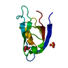
| ||||||||
|---|---|---|---|---|---|---|---|---|---|
| 1 |
| ||||||||
| Unit cell |
|
-Components
| #1: Protein | Synapse / Synapse-associated protein 102 / Neuroendocrine-DLG / NE-DLG / Discs large homolog 3 / DLG3 / DLG3A Mass: 9972.403 Da / Num. of mol.: 1 / Fragment: PDZ2 Source method: isolated from a genetically manipulated source Source: (gene. exp.) Homo sapiens (human) / Gene: DLG3, KIAA1232 / Plasmid: pNIC28-Bsa4 / Production host:  Escherichia coli (E. coli) / Strain (production host): BL21(DE3)-R3 / References: UniProt: Q92796 Escherichia coli (E. coli) / Strain (production host): BL21(DE3)-R3 / References: UniProt: Q92796 |
|---|---|
| #2: Chemical | ChemComp-SO4 / Sulfate  Mass: 96.063 Da / Num. of mol.: 1 / Source method: obtained synthetically / Formula: SO4 Mass: 96.063 Da / Num. of mol.: 1 / Source method: obtained synthetically / Formula: SO4 |
| #3: Chemical | ChemComp-GOL / Glycerol  Mass: 92.094 Da / Num. of mol.: 1 / Source method: obtained synthetically / Formula: C3H8O3 Mass: 92.094 Da / Num. of mol.: 1 / Source method: obtained synthetically / Formula: C3H8O3 |
| #4: Water | ChemComp-HOH / Water Mass: 18.015 Da / Num. of mol.: 173 / Source method: isolated from a natural source / Formula: H2O Mass: 18.015 Da / Num. of mol.: 173 / Source method: isolated from a natural source / Formula: H2O |
-Experimental details
-Experiment
| Experiment | Method: X-RAY DIFFRACTION / Number of used crystals: 1 |
|---|
- Sample preparation
| Crystal | Density Matthews: 1.78 Å3/Da / Density % sol: 30.88 % |
|---|---|
| Crystal grow | Temperature: 293 K / Method: vapor diffusion, sitting drop / pH: 5.5 Details: Ammonium sulphate, Glycerol, Bis-Tris, pH 5.5, VAPOR DIFFUSION, SITTING DROP, temperature 293K |
-Data collection
| Diffraction | Mean temperature: 100 K |
|---|---|
| Diffraction source | Source: SYNCHROTRON / Site: SLS  / Beamline: X10SA / Wavelength: 0.9791 Å / Beamline: X10SA / Wavelength: 0.9791 Å |
| Detector | Type: MARMOSAIC 225 mm CCD / Detector: CCD / Date: Nov 18, 2005 |
| Radiation | Protocol: SINGLE WAVELENGTH / Monochromatic (M) / Laue (L): M / Scattering type: x-ray |
| Radiation wavelength | Wavelength: 0.9791 Å / Relative weight: 1 |
| Reflection | Resolution: 1.1→26.45 Å / Num. obs: 27971 / % possible obs: 98.4 % / Observed criterion σ(F): 0 / Observed criterion σ(I): 0 |
| Reflection shell | Resolution: 1.1→1.16 Å / % possible all: 96.6 |
- Processing
| Software |
| ||||||||||||||||||||||||||||||||||||||||||||||||||||||||||||||||||||||||||||||||||||||||||||||||||||||||||||||||||||||||||||||||||||||||||||
|---|---|---|---|---|---|---|---|---|---|---|---|---|---|---|---|---|---|---|---|---|---|---|---|---|---|---|---|---|---|---|---|---|---|---|---|---|---|---|---|---|---|---|---|---|---|---|---|---|---|---|---|---|---|---|---|---|---|---|---|---|---|---|---|---|---|---|---|---|---|---|---|---|---|---|---|---|---|---|---|---|---|---|---|---|---|---|---|---|---|---|---|---|---|---|---|---|---|---|---|---|---|---|---|---|---|---|---|---|---|---|---|---|---|---|---|---|---|---|---|---|---|---|---|---|---|---|---|---|---|---|---|---|---|---|---|---|---|---|---|---|---|
| Refinement | Method to determine structure: MOLECULAR REPLACEMENT Starting model: 2BYG.pdb Resolution: 1.1→26.45 Å / Cor.coef. Fo:Fc: 0.981 / Cor.coef. Fo:Fc free: 0.97 / SU B: 0.886 / SU ML: 0.02 / Cross valid method: THROUGHOUT / σ(F): 0 / ESU R: 0.029 / ESU R Free: 0.032 / Stereochemistry target values: MAXIMUM LIKELIHOOD / Details: HYDROGENS HAVE BEEN ADDED IN THE RIDING POSITIONS
| ||||||||||||||||||||||||||||||||||||||||||||||||||||||||||||||||||||||||||||||||||||||||||||||||||||||||||||||||||||||||||||||||||||||||||||
| Solvent computation | Ion probe radii: 0.8 Å / Shrinkage radii: 0.8 Å / VDW probe radii: 1.4 Å / Solvent model: BABINET MODEL WITH MASK | ||||||||||||||||||||||||||||||||||||||||||||||||||||||||||||||||||||||||||||||||||||||||||||||||||||||||||||||||||||||||||||||||||||||||||||
| Displacement parameters | Biso mean: 7.076 Å2
| ||||||||||||||||||||||||||||||||||||||||||||||||||||||||||||||||||||||||||||||||||||||||||||||||||||||||||||||||||||||||||||||||||||||||||||
| Refinement step | Cycle: LAST / Resolution: 1.1→26.45 Å
| ||||||||||||||||||||||||||||||||||||||||||||||||||||||||||||||||||||||||||||||||||||||||||||||||||||||||||||||||||||||||||||||||||||||||||||
| Refine LS restraints |
| ||||||||||||||||||||||||||||||||||||||||||||||||||||||||||||||||||||||||||||||||||||||||||||||||||||||||||||||||||||||||||||||||||||||||||||
| LS refinement shell | Resolution: 1.1→1.129 Å / Total num. of bins used: 20
|
