 Movie
Movie Controller

[English] 日本語
 Yorodumi
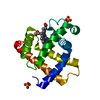
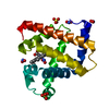
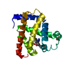
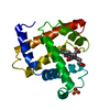
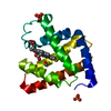
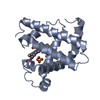
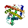
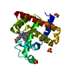
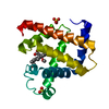
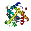
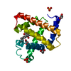
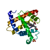
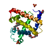
- PDB-2ekt: Crystal structure of myoglobin reconstituted with 6-methyl-6-depr... -
+ Open data

- Basic information
| Entry | Database: PDB / ID: 2ekt | ||||||
|---|---|---|---|---|---|---|---|
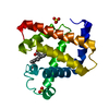
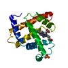
| Title | Crystal structure of myoglobin reconstituted with 6-methyl-6-depropionatehemin | ||||||
 Components Components | Myoglobin | ||||||
 Keywords Keywords | OXYGEN STORAGE/TRANSPORT / globin fold / OXYGEN STORAGE-TRANSPORT COMPLEX | ||||||
| Function / homology |  Function and homology information Function and homology informationhydrogen peroxide mediated signaling pathway / oxygen carrier activity / oxygen binding / heme binding / metal ion bindingSimilarity search - Function | ||||||
| Biological species |  Physeter catodon (sperm whale) Physeter catodon (sperm whale) | ||||||
| Method | X-RAY DIFFRACTION / SYNCHROTRON / MOLECULAR REPLACEMENT / Resolution: 1.1 Å | ||||||
 Authors Authors | Harada, K. / Makino, M. / Sugimoto, H. / Hirota, S. / Matsuo, T. / Shiro, Y. / Hisaeda, Y. / Hayashi, T. | ||||||
 Citation Citation | Journal: Biochemistry / Year: 2007 Title: Structure and ligand binding properties of myoglobins reconstituted with monodepropionated heme: functional role of each heme propionate side chain Authors: Harada, K. / Makino, M. / Sugimoto, H. / Hirota, S. / Matsuo, T. / Shiro, Y. / Hisaeda, Y. / Hayashi, T. | ||||||
| History |
|
- Structure visualization
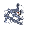
| Structure viewer | Molecule: MolmilJmol/JSmol |
|---|
- Downloads & links
-Download
| PDBx/mmCIF format | 2ekt.cif.gz | 93.2 KB | Display | PDBx/mmCIF format |
|---|---|---|---|---|
| PDB format | pdb2ekt.ent.gz | 69.8 KB | Display | PDB format |
| PDBx/mmJSON format | 2ekt.json.gz | Tree view | PDBx/mmJSON format | |
| Others |  Other downloads Other downloads |
-Validation report
| Arichive directory | https://data.pdbj.org/pub/pdb/validation_reports/ek/2ektftp://data.pdbj.org/pub/pdb/validation_reports/ek/2ekt | HTTPS FTP |
|---|
-Related structure data
| Related structure data | 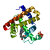 2ekuC 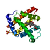 1jw8S C: citing same article ( S: Starting model for refinement |
|---|---|
| Similar structure data |


-Links
 PDBj
PDBj


- Assembly
| Deposited unit | 
| ||||||||
|---|---|---|---|---|---|---|---|---|---|
| 1 |
| ||||||||
| Unit cell |
| ||||||||
| Components on special symmetry positions |
|
-Components
| #1: Protein | Mass: 17234.951 Da / Num. of mol.: 1 / Source method: isolated from a natural source / Source: (natural) Physeter catodon (sperm whale) / Tissue: Skeletal Muscle / References: UniProt: P02185 | ||||
|---|---|---|---|---|---|
| #2: Chemical | ChemComp-SO4 / Sulfate  Mass: 96.063 Da / Num. of mol.: 4 / Source method: obtained synthetically / Formula: SO4 Mass: 96.063 Da / Num. of mol.: 4 / Source method: obtained synthetically / Formula: SO4#3: Chemical | ChemComp-6HE / | 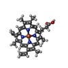  Mass: 558.451 Da / Num. of mol.: 1 / Source method: obtained synthetically / Formula: C32H30FeN4O2 Mass: 558.451 Da / Num. of mol.: 1 / Source method: obtained synthetically / Formula: C32H30FeN4O2#4: Water | ChemComp-HOH / | Water Mass: 18.015 Da / Num. of mol.: 303 / Source method: isolated from a natural source / Formula: H2O Mass: 18.015 Da / Num. of mol.: 303 / Source method: isolated from a natural source / Formula: H2O |
-Experimental details
-Experiment
| Experiment | Method: X-RAY DIFFRACTION / Number of used crystals: 1 |
|---|
- Sample preparation
| Crystal | Density Matthews: 3.09 Å3/Da / Density % sol: 60.22 % |
|---|---|
| Crystal grow | Temperature: 277 K / Method: vapor diffusion, sitting drop / pH: 6.4 Details: 3.0M ammonium sulfate, 100mM bis-tris propane, pH 6.4, VAPOR DIFFUSION, SITTING DROP, temperature 277K |
-Data collection
| Diffraction | Mean temperature: 100 K |
|---|---|
| Diffraction source | Source: SYNCHROTRON / Site: SPring-8  / Beamline: BL44B2 / Wavelength: 0.78 Å / Beamline: BL44B2 / Wavelength: 0.78 Å |
| Detector | Type: ADSC QUANTUM 210 / Detector: CCD / Date: Feb 18, 2004 |
| Radiation | Monochromator: SI(III) MIRRORS / Protocol: SINGLE WAVELENGTH / Monochromatic (M) / Laue (L): M / Scattering type: x-ray |
| Radiation wavelength | Wavelength: 0.78 Å / Relative weight: 1 |
| Reflection | Resolution: 1.1→50 Å / Num. obs: 85421 / % possible obs: 100 % / Observed criterion σ(I): 0 / Redundancy: 23.5 % / Rsym value: 0.062 / Net I/σ(I): 38.7 |
| Reflection shell | Resolution: 1.1→1.14 Å / Redundancy: 14.6 % / Mean I/σ(I) obs: 6.2 / Num. unique all: 8466 / Rsym value: 0.349 / % possible all: 100 |
- Processing
| Software |
| |||||||||||||||||||||||||||||||||
|---|---|---|---|---|---|---|---|---|---|---|---|---|---|---|---|---|---|---|---|---|---|---|---|---|---|---|---|---|---|---|---|---|---|---|
| Refinement | Method to determine structure: MOLECULAR REPLACEMENT Starting model: PDB ENTRY 1JW8 Resolution: 1.1→10 Å / Num. parameters: 14631 / Num. restraintsaints: 17821 / Cross valid method: THROUGHOUT / σ(F): 0 / σ(I): 0 / Stereochemistry target values: ENGH & HUBER
| |||||||||||||||||||||||||||||||||
| Refine analyze | Num. disordered residues: 5 / Occupancy sum hydrogen: 1095 / Occupancy sum non hydrogen: 1577.33 | |||||||||||||||||||||||||||||||||
| Refinement step | Cycle: LAST / Resolution: 1.1→10 Å
| |||||||||||||||||||||||||||||||||
| Refine LS restraints |
|
