 Movie
Movie Controller

[English] 日本語
 Yorodumi
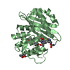
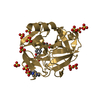
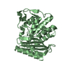
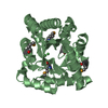
- PDB-2ei5: Crystal Structure of Hypothetical protein(TTHA0061) from Thermus ... -
+ Open data

- Basic information
| Entry | Database: PDB / ID: 2ei5 | ||||||
|---|---|---|---|---|---|---|---|
| Title | Crystal Structure of Hypothetical protein(TTHA0061) from Thermus thermophilus | ||||||
 Components Components | Hypothetical protein TTHA0061 Hypothesis Hypothesis | ||||||
 Keywords Keywords | STRUCTURAL GENOMICS / UNKNOWN FUNCTION / Hypothetical protein / Thermus thermophilus / TTHA0061 / NPPSFA / National Project on Protein Structural and Functional Analyses / RIKEN Structural Genomics/Proteomics Initiative / RSGI | ||||||
| Function / homology | Protein of unknown function (DUF3208) / Protein of unknown function DUF3208 / Protein of unknown function (DUF3208) / Alpha-Beta Plaits / 2-Layer Sandwich / Alpha Beta / DUF3208 domain-containing protein Function and homology information Function and homology information | ||||||
| Biological species |   Thermus thermophilus (bacteria) Thermus thermophilus (bacteria) | ||||||
| Method | X-RAY DIFFRACTION / SYNCHROTRON / MOLECULAR REPLACEMENT / Resolution: 1.88 Å | ||||||
 Authors Authors | Niwa, H. / Kumarevel, T.S. / Kuramitsu, S. / Yokoyama, S. / RIKEN Structural Genomics/Proteomics Initiative (RSGI) | ||||||
 Citation Citation | Journal: To be Published Title: Crystal Structure of Hypothetical protein(TTHA0061) from Thermus thermophilus Authors: Niwa, H. / Kumarevel, T.S. / Kuramitsu, S. / Yokoyama, S. | ||||||
| History |
|
- Structure visualization
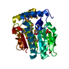
| Structure viewer | Molecule: MolmilJmol/JSmol |
|---|
- Downloads & links
-Download
| PDBx/mmCIF format | 2ei5.cif.gz | 56.8 KB | Display | PDBx/mmCIF format |
|---|---|---|---|---|
| PDB format | pdb2ei5.ent.gz | 41.2 KB | Display | PDB format |
| PDBx/mmJSON format | 2ei5.json.gz | Tree view | PDBx/mmJSON format | |
| Others |  Other downloads Other downloads |
-Validation report
| Arichive directory | https://data.pdbj.org/pub/pdb/validation_reports/ei/2ei5ftp://data.pdbj.org/pub/pdb/validation_reports/ei/2ei5 | HTTPS FTP |
|---|
-Related structure data
| Related structure data |  2ebgS S: Starting model for refinement |
|---|---|
| Similar structure data | |
| Other databases |


-Links
 PDBj
PDBj- Assembly
| Deposited unit | 
| ||||||||
|---|---|---|---|---|---|---|---|---|---|
| 1 |
| ||||||||
| Unit cell |
|
-Components
| #1: Protein | Hypothesis Mass: 11842.597 Da / Num. of mol.: 2 Source method: isolated from a genetically manipulated source Source: (gene. exp.) Thermus thermophilus (bacteria) / Gene: TTHA0061 / Plasmid: pET-11a / Species (production host): Escherichia coli / Production host: Escherichia coli BL21(DE3) (bacteria) / Strain (production host): BL21(DE3) / References: UniProt: Q5SM82#2: Chemical | ChemComp-BTB / | Bis-tris methane  Mass: 209.240 Da / Num. of mol.: 1 / Source method: obtained synthetically / Formula: C8H19NO5 / Comment: pH buffer*YM Mass: 209.240 Da / Num. of mol.: 1 / Source method: obtained synthetically / Formula: C8H19NO5 / Comment: pH buffer*YM#3: Water | ChemComp-HOH / | Water Mass: 18.015 Da / Num. of mol.: 150 / Source method: isolated from a natural source / Formula: H2O Mass: 18.015 Da / Num. of mol.: 150 / Source method: isolated from a natural source / Formula: H2O |
|---|
-Experimental details
-Experiment
| Experiment | Method: X-RAY DIFFRACTION / Number of used crystals: 1 |
|---|
- Sample preparation
| Crystal | Density Matthews: 2.12 Å3/Da / Density % sol: 42 % |
|---|---|
| Crystal grow | Temperature: 293 K / Method: vapor diffusion, sitting drop / pH: 6 Details: 28% PEG MME2000, 0.1M Bis-Tris pH6.0, VAPOR DIFFUSION, SITTING DROP, temperature 293K |
-Data collection
| Diffraction | Mean temperature: 100 K |
|---|---|
| Diffraction source | Source: SYNCHROTRON / Site: SPring-8  / Beamline: BL26B2 / Wavelength: 1 Å / Beamline: BL26B2 / Wavelength: 1 Å |
| Detector | Type: RIGAKU JUPITER 210 / Detector: CCD / Date: Sep 30, 2005 |
| Radiation | Monochromator: Si / Protocol: SINGLE WAVELENGTH / Monochromatic (M) / Laue (L): M / Scattering type: x-ray |
| Radiation wavelength | Wavelength: 1 Å / Relative weight: 1 |
| Reflection | Resolution: 1.88→50 Å / Num. all: 15964 / Num. obs: 15964 / % possible obs: 99.8 % / Observed criterion σ(F): 2 / Observed criterion σ(I): 0 / Redundancy: 10.2 % / Biso Wilson estimate: 21 Å2 / Rmerge(I) obs: 0.038 |
| Reflection shell | Resolution: 1.88→1.95 Å / Redundancy: 7.8 % / Rmerge(I) obs: 0.239 / Num. unique all: 1580 / % possible all: 98.9 |
- Processing
| Software |
| ||||||||||||||||||||||||||||||||||||
|---|---|---|---|---|---|---|---|---|---|---|---|---|---|---|---|---|---|---|---|---|---|---|---|---|---|---|---|---|---|---|---|---|---|---|---|---|---|
| Refinement | Method to determine structure: MOLECULAR REPLACEMENT Starting model: 2EBG Resolution: 1.88→41.65 Å / Rfactor Rfree error: 0.009 / Data cutoff high absF: 1047770.47 / Data cutoff low absF: 0 / Isotropic thermal model: RESTRAINED / Cross valid method: THROUGHOUT / σ(F): 0 / Stereochemistry target values: Engh & Huber
| ||||||||||||||||||||||||||||||||||||
| Solvent computation | Solvent model: FLAT MODEL / Bsol: 39.7388 Å2 / ksol: 0.328653 e/Å3 | ||||||||||||||||||||||||||||||||||||
| Displacement parameters | Biso mean: 33.5 Å2
| ||||||||||||||||||||||||||||||||||||
| Refine analyze |
| ||||||||||||||||||||||||||||||||||||
| Refinement step | Cycle: LAST / Resolution: 1.88→41.65 Å
| ||||||||||||||||||||||||||||||||||||
| Refine LS restraints |
| ||||||||||||||||||||||||||||||||||||
| LS refinement shell | Resolution: 1.88→2 Å / Rfactor Rfree error: 0.022 / Total num. of bins used: 6
| ||||||||||||||||||||||||||||||||||||
| Xplor file |
|
