 Movie
Movie Controller


+ Open data

- Basic information
| Entry | Database: PDB / ID: 2edg | ||||||
|---|---|---|---|---|---|---|---|
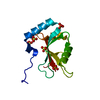
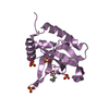
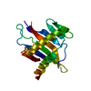
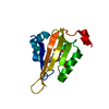
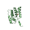
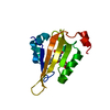
| Title | Solution structure of the GCV_H domain from mouse glycine | ||||||
 Components Components | Glycine cleavage system H protein | ||||||
 Keywords Keywords | BIOSYNTHETIC PROTEIN / Barrel-sandwich hybrid / Structural Genomics / NPPSFA / National Project on Protein Structural and Functional Analyses / RIKEN Structural Genomics/Proteomics Initiative / RSGI | ||||||
| Function / homology |  Function and homology information Function and homology informationGlycine degradation / aminomethyltransferase activity / glycine cleavage complex / glycine decarboxylation via glycine cleavage system / Glyoxylate metabolism and glycine degradation / protein lipoylation / enzyme binding / mitochondrionSimilarity search - Function | ||||||
| Biological species |  Mus musculus (house mouse) Mus musculus (house mouse) | ||||||
| Method | SOLUTION NMR / torsion angle dynamics | ||||||
 Authors Authors | Endo, H. / Yoshida, M. / Hayashi, F. / Yokoyama, S. / RIKEN Structural Genomics/Proteomics Initiative (RSGI) | ||||||
 Citation Citation | Journal: To be Published Title: Solution structure of the GCV_H domain from mouse glycine Authors: Endo, H. / Yoshida, M. / Hayashi, F. / Yokoyama, S. | ||||||
| History |
|
- Structure visualization
| Structure viewer | Molecule: MolmilJmol/JSmol |
|---|
- Downloads & links
-Download
| PDBx/mmCIF format | 2edg.cif.gz | 748.8 KB | Display | PDBx/mmCIF format |
|---|---|---|---|---|
| PDB format | pdb2edg.ent.gz | 651.6 KB | Display | PDB format |
| PDBx/mmJSON format | 2edg.json.gz | Tree view | PDBx/mmJSON format | |
| Others |  Other downloads Other downloads |
-Validation report
| Arichive directory | https://data.pdbj.org/pub/pdb/validation_reports/ed/2edgftp://data.pdbj.org/pub/pdb/validation_reports/ed/2edg | HTTPS FTP |
|---|
-Related structure data
| Similar structure data | |
|---|---|
| Other databases |


-Links
 PDBj
PDBj

- Assembly
| Deposited unit | 
| |||||||||
|---|---|---|---|---|---|---|---|---|---|---|
| 1 |
| |||||||||
| NMR ensembles |
|
-Components
| #1: Protein | Mass: 14188.617 Da / Num. of mol.: 1 / Fragment: GCV_H Source method: isolated from a genetically manipulated source Source: (gene. exp.) Mus musculus (house mouse) / Description: cell free protein synthesis / Gene: Gcsh / Plasmid: P060313-07 / References: UniProt: Q91WK5 |
|---|
-Experimental details
-Experiment
| Experiment | Method: SOLUTION NMR | ||||||||||||
|---|---|---|---|---|---|---|---|---|---|---|---|---|---|
| NMR experiment |
|
- Sample preparation
| Details | Contents: 0.79mM 13C, 15N-labeled protein; 20mM d-Tris-HCl(pH 7.0); 100mM NaCl; 1mM d-DTT; 0.02% NaN3; 10% D2O, 90% H2O Solvent system: 10% D2O, 90% H2O |
|---|---|
| Sample conditions | Ionic strength: 120mM / pH: 7.0 / Pressure: ambient / Temperature: 298 K |
-NMR measurement
| NMR spectrometer | Type: Varian INOVA / Manufacturer: Varian / Model: INOVA / Field strength: 800 MHz |
|---|
- Processing
| NMR software |
| ||||||||||||||||||||||||||||
|---|---|---|---|---|---|---|---|---|---|---|---|---|---|---|---|---|---|---|---|---|---|---|---|---|---|---|---|---|---|
| Refinement | Method: torsion angle dynamics / Software ordinal: 1 | ||||||||||||||||||||||||||||
| NMR representative | Selection criteria: lowest energy | ||||||||||||||||||||||||||||
| NMR ensemble | Conformer selection criteria: structures with the least restraint violations, target function Conformers calculated total number: 100 / Conformers submitted total number: 20 |
