 Movie
Movie Controller

[English] 日本語
 Yorodumi
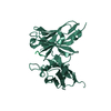
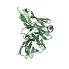
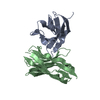
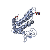
- PDB-2dlf: HIGH RESOLUTION CRYSTAL STRUCTURE OF THE FV FRAGMENT FROM AN ANTI... -
+ Open data

- Basic information
| Entry | Database: PDB / ID: 2dlf | ||||||
|---|---|---|---|---|---|---|---|
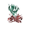
| Title | HIGH RESOLUTION CRYSTAL STRUCTURE OF THE FV FRAGMENT FROM AN ANTI-DANSYL SWITCH VARIANT ANTIBODY IGG2A(S) CRYSTALLIZED AT PH 6.75 | ||||||
 Components Components |
| ||||||
 Keywords Keywords |  IMMUNE SYSTEM / FV FRAGMENT / IMMUNOGLOBULIN IMMUNE SYSTEM / FV FRAGMENT / IMMUNOGLOBULIN | ||||||
| Function / homology |  Function and homology informationimmunoglobulin complex / immunoglobulin mediated immune response / antigen binding / adaptive immune response / immune response / extracellular space Function and homology informationimmunoglobulin complex / immunoglobulin mediated immune response / antigen binding / adaptive immune response / immune response / extracellular spaceSimilarity search - Function | ||||||
| Biological species |  Mus musculus (house mouse) Mus musculus (house mouse) | ||||||
| Method | X-RAY DIFFRACTION / MOLECULAR REPLACEMENT / Resolution: 1.55 Å | ||||||
 Authors Authors | Nakasako, M. / Takahashi, H. / Shimada, I. / Arata, Y. | ||||||
 Citation Citation | Journal: J.Mol.Biol. / Year: 1999 Title: The pH-dependent structural variation of complementarity-determining region H3 in the crystal structures of the Fv fragment from an anti-dansyl monoclonal antibody. Authors: Nakasako, M. / Takahashi, H. / Shimba, N. / Shimada, I. / Arata, Y. #1: Journal: J.Mol.Biol. / Year: 1994Title: Role of the Domain-Domain Interaction in the Construction of the Antigen Combining Site. A Comparative Study by 1H-15N Shift Correlation NMR Spectroscopy of the Fv and Fab Fragment of Anti- ...Title: Role of the Domain-Domain Interaction in the Construction of the Antigen Combining Site. A Comparative Study by 1H-15N Shift Correlation NMR Spectroscopy of the Fv and Fab Fragment of Anti-Dansyl Mouse Monoclonal Antibody Authors: Takahashi, H. / Tamura, H. / Shimba, N. / Shimada, I. / Arata, Y. #2: Journal: Biochemistry / Year: 1992Title: Dynamical Structure of the Antibody Combining Site as Studied by 1H-15N Shift Correlation NMR Spectroscopy Authors: Takahashi, H. / Suzuki, E. / Ishimadaarata, Y. #3: Journal: Biochemistry / Year: 1992Title: Isotope-Edited Nuclear Magnetic Resonance Study of Fv Fragment of an Anti- Dansyl Mouse Monoclonal Antibody: Recognition of the Dansyl Hapten Authors: Odaka, A. / Kim, J.I. / Takahashi, H. / Shimada, I. / Arata, Y. #4: Journal: Biochemistry / Year: 1991Title: Preparation of the Fv Fragment from a Short-Chain Mouse IgG2a Anti-Dansyl Monoclonal Antibody and Use of Selectively Deuterated Fv Analogues for Two- Dimensional 1H NMR Analyses of Antigen-Antibody Interactions Authors: Takahashi, H. / Igarashi, T. / Shimada, I. / Arata, Y. #5: Journal: Biochemistry / Year: 1991Title: Multinuclear NMR Study of the Structure of the Fv Fragment of Anti-Dansyl Mouse IgG2a Antibody Authors: Takahashi, H. / Odaka, A. / Kawaminami, S. / Matsunaga, C. / Kato, K. / Shimada, I. / Arata, Y. #6: Journal: J.Am.Chem.Soc. / Year: 1991Title: Conformation and Stereoselective Reduction of Hapten Side Chain in the Antibody Combining Site Authors: Kimnakano, T. / Higuchi, T. / Hirobe, M. / Shimada, I. / Arata, Y. #7: Journal: Biochemistry / Year: 1990Title: Structure of a Mouse Immunoglobulin G that Lacks the Entire CH1 Domain: Protein Sequencing and Small-Angle X-Ray Scattering Studies Authors: Igarashi, T. / Sato, M. / Katsube, Y. / Takio, K. / Tanaka, T. / Nakanishi, M. / Arata, Y. | ||||||
| History |
|
- Structure visualization
| Structure viewer | Molecule: MolmilJmol/JSmol |
|---|
- Downloads & links
-Download
| PDBx/mmCIF format | 2dlf.cif.gz | 71.7 KB | Display | PDBx/mmCIF format |
|---|---|---|---|---|
| PDB format | pdb2dlf.ent.gz | 51.1 KB | Display | PDB format |
| PDBx/mmJSON format | 2dlf.json.gz | Tree view | PDBx/mmJSON format | |
| Others |  Other downloads Other downloads |
-Validation report
| Arichive directory | https://data.pdbj.org/pub/pdb/validation_reports/dl/2dlfftp://data.pdbj.org/pub/pdb/validation_reports/dl/2dlf | HTTPS FTP |
|---|
-Related structure data
| Related structure data |  1dlfC  1igfS S: Starting model for refinement C: citing same article ( |
|---|---|
| Similar structure data |


-Links
 PDBj
PDBj

- Assembly
| Deposited unit | 
| ||||||||
|---|---|---|---|---|---|---|---|---|---|
| 1 |
| ||||||||
| Unit cell |
|
-Components
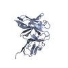
| #1: Antibody | Mass: 12386.949 Da / Num. of mol.: 1 Fragment: FV FRAGMENT (VL DOMAIN) SYNONYM: ANTI-DANSYL FV FRAGMENT Source method: isolated from a natural source / Source: (natural) Mus musculus (house mouse) / Cell line: HYBRIDOMA / References: UniProt: P01631 | ||||
|---|---|---|---|---|---|
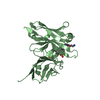
| #2: Antibody | Mass: 14152.787 Da / Num. of mol.: 1 Fragment: FV FRAGMENT (VH DOMAIN) SYNONYM: ANTI-DANSYL FV FRAGMENT Source method: isolated from a natural source / Source: (natural) Mus musculus (house mouse) / Cell line: HYBRIDOMA / References: UniProt: P01801 | ||||
| #3: Chemical | Sulfate  Mass: 96.063 Da / Num. of mol.: 2 / Source method: obtained synthetically / Formula: SO4 Mass: 96.063 Da / Num. of mol.: 2 / Source method: obtained synthetically / Formula: SO4#4: Water | ChemComp-HOH / | Water Mass: 18.015 Da / Num. of mol.: 400 / Source method: isolated from a natural source / Formula: H2O Mass: 18.015 Da / Num. of mol.: 400 / Source method: isolated from a natural source / Formula: H2OCompound details | COMPAOUND THE FV FRAGMENT WAS PROPARED BY A PROTEOLYTIC DIGESTION OF ANTI-DANSYL MONOCLONAL ...COMPAOUND THE FV FRAGMENT WAS PROPARED BY A PROTEOLYTI | |
-Experimental details
-Experiment
| Experiment | Method: X-RAY DIFFRACTION / Number of used crystals: 1 |
|---|
- Sample preparation
| Crystal | Density Matthews: 1.96 Å3/Da / Density % sol: 37.7 % / Description: DATA WERE COLLECTED BY OSCILLATION METHOD | ||||||||||||||||||||||||||||||
|---|---|---|---|---|---|---|---|---|---|---|---|---|---|---|---|---|---|---|---|---|---|---|---|---|---|---|---|---|---|---|---|
| Crystal grow | Method: vapor diffusion, hanging drop / pH: 6.75 Details: CRYSTALLIZATION CONDITIONS: TEMPERATURE AT 20C. PRECIPITANT SOLUTION CONTAINED 1 AMMONIUM SULFATE AND 0.025 M SODIUM CACODYLATE (PH 6.75). 5 MG/ML OF FV FRAGMENT., VAPOR DIFFUSION, HANGING DROP | ||||||||||||||||||||||||||||||
| Crystal grow | *PLUS Temperature: 293 K | ||||||||||||||||||||||||||||||
| Components of the solutions | *PLUS
|
-Data collection
| Diffraction | Mean temperature: 112 K |
|---|---|
| Diffraction source | Source: ROTATING ANODE / Type: RIGAKU ULTRAX 18 / Wavelength: 1.5418 |
| Detector | Type: RIGAKU RAXIS IV / Detector: IMAGE PLATE / Date: Jul 10, 1998 / Details: DOUBLE FOCUSING MIRROR (PT COATED AND NI COATED) |
| Radiation | Monochromator: NI FILTER / Protocol: SINGLE WAVELENGTH / Monochromatic (M) / Laue (L): M / Scattering type: x-ray |
| Radiation wavelength | Wavelength: 1.5418 Å / Relative weight: 1 |
| Reflection | Resolution: 1.5→40 Å / Num. obs: 32405 / % possible obs: 97.6 % / Observed criterion σ(I): 1 / Redundancy: 11.1 % / Rmerge(I) obs: 0.053 / Net I/σ(I): 42.6 |
| Reflection shell | Resolution: 1.5→1.52 Å / Redundancy: 5.4 % / Rmerge(I) obs: 0.318 / Mean I/σ(I) obs: 3.5 / % possible all: 93 |
| Reflection | *PLUS Highest resolution: 1.55 Å / Num. obs: 29973 / % possible obs: 97.7 % / Num. measured all: 331744 / Rmerge(I) obs: 0.052 |
| Reflection shell | *PLUS Highest resolution: 1.55 Å / Lowest resolution: 1.57 Å / % possible obs: 96.3 % / Mean I/σ(I) obs: 4.5 |
- Processing
| Software |
| ||||||||||||||||||||||||||||||||||||||||||||||||||||||||||||
|---|---|---|---|---|---|---|---|---|---|---|---|---|---|---|---|---|---|---|---|---|---|---|---|---|---|---|---|---|---|---|---|---|---|---|---|---|---|---|---|---|---|---|---|---|---|---|---|---|---|---|---|---|---|---|---|---|---|---|---|---|---|
| Refinement | Method to determine structure: MOLECULAR REPLACEMENT Starting model: 1IGF Resolution: 1.55→8 Å / Rfactor Rfree error: 0.004 / Data cutoff high absF: 1000 / Data cutoff low absF: 1 / Isotropic thermal model: GAUSS / Cross valid method: THROUGHOUT / σ(F): 2 / Details: ALL ATOMS IN THE ASYMMETRIC UNIT WERE REFINED
| ||||||||||||||||||||||||||||||||||||||||||||||||||||||||||||
| Displacement parameters | Biso mean: 13.8 Å2 | ||||||||||||||||||||||||||||||||||||||||||||||||||||||||||||
| Refine analyze | Luzzati coordinate error obs: 0.17 Å | ||||||||||||||||||||||||||||||||||||||||||||||||||||||||||||
| Refinement step | Cycle: LAST / Resolution: 1.55→8 Å
| ||||||||||||||||||||||||||||||||||||||||||||||||||||||||||||
| Refine LS restraints |
| ||||||||||||||||||||||||||||||||||||||||||||||||||||||||||||
| LS refinement shell | Resolution: 1.55→1.57 Å / Rfactor Rfree error: 0.037 / Total num. of bins used: 26
| ||||||||||||||||||||||||||||||||||||||||||||||||||||||||||||
| Xplor file | Serial no: 1 / Param file: PARHCSDX.PRO / Topol file: TOPHCSDX.PRO | ||||||||||||||||||||||||||||||||||||||||||||||||||||||||||||
| Software | *PLUS Name: X-PLOR / Version: 3.1 / Classification: refinement | ||||||||||||||||||||||||||||||||||||||||||||||||||||||||||||
| Refinement | *PLUS Num. reflection obs: 28800 / Rfactor obs: 0.179 / Rfactor Rfree: 0.228 / Rfactor Rwork: 0.179 | ||||||||||||||||||||||||||||||||||||||||||||||||||||||||||||
| Solvent computation | *PLUS | ||||||||||||||||||||||||||||||||||||||||||||||||||||||||||||
| Displacement parameters | *PLUS | ||||||||||||||||||||||||||||||||||||||||||||||||||||||||||||
| Refine LS restraints | *PLUS
|
