 Movie
Movie Controller

[English] 日本語
 Yorodumi
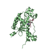
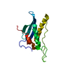
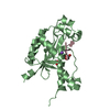
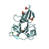
- PDB-2cyx: Structure of human ubiquitin-conjugating enzyme E2 G2 (UBE2G2/UBC7) -
+ Open data

- Basic information
| Entry | Database: PDB / ID: 2cyx | ||||||
|---|---|---|---|---|---|---|---|
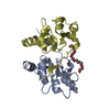
| Title | Structure of human ubiquitin-conjugating enzyme E2 G2 (UBE2G2/UBC7) | ||||||
 Components Components | Ubiquitin-conjugating enzyme E2 G2 | ||||||
 Keywords Keywords |  LIGASE / Ubiquitin-conjugating enzyme (E2) / Ubl conjugation pathway / Structural Genomics / NPPSFA / National Project on Protein Structural and Functional Analyses / RIKEN Structural Genomics/Proteomics Initiative / RSGI LIGASE / Ubiquitin-conjugating enzyme (E2) / Ubl conjugation pathway / Structural Genomics / NPPSFA / National Project on Protein Structural and Functional Analyses / RIKEN Structural Genomics/Proteomics Initiative / RSGI | ||||||
| Function / homology |  Function and homology information Function and homology informationnegative regulation of retrograde protein transport, ER to cytosol / E2 ubiquitin-conjugating enzyme / ubiquitin conjugating enzyme activity / protein K48-linked ubiquitination / cellular response to interferon-beta / : / Synthesis of active ubiquitin: roles of E1 and E2 enzymes / lipid droplet / ubiquitin-protein transferase activity / Antigen processing: Ubiquitination & Proteasome degradation ...negative regulation of retrograde protein transport, ER to cytosol / E2 ubiquitin-conjugating enzyme / ubiquitin conjugating enzyme activity / protein K48-linked ubiquitination / cellular response to interferon-beta / : / Synthesis of active ubiquitin: roles of E1 and E2 enzymes / lipid droplet / ubiquitin-protein transferase activity / Antigen processing: Ubiquitination & Proteasome degradation / ubiquitin-dependent protein catabolic process / endoplasmic reticulum / ATP binding / identical protein binding / cytosolSimilarity search - Function | ||||||
| Biological species |  Homo sapiens (human) Homo sapiens (human) | ||||||
| Method | X-RAY DIFFRACTION / SYNCHROTRON / MOLECULAR REPLACEMENT / Resolution: 2.56 Å | ||||||
 Authors Authors | Yoshikawa, S. / Arai, R. / Murayama, K. / Imai, Y. / Takahashi, R. / Shirouzu, M. / Yokoyama, S. / RIKEN Structural Genomics/Proteomics Initiative (RSGI) | ||||||
 Citation Citation | Journal: Acta Crystallogr.,Sect.F / Year: 2006 Title: Structure of human ubiquitin-conjugating enzyme E2 G2 (UBE2G2/UBC7) Authors: Arai, R. / Yoshikawa, S. / Murayama, K. / Imai, Y. / Takahashi, R. / Shirouzu, M. / Yokoyama, S. | ||||||
| History |
|
- Structure visualization
| Structure viewer | Molecule: MolmilJmol/JSmol |
|---|
- Downloads & links
-Download
| PDBx/mmCIF format | 2cyx.cif.gz | 110.4 KB | Display | PDBx/mmCIF format |
|---|---|---|---|---|
| PDB format | pdb2cyx.ent.gz | 86.1 KB | Display | PDB format |
| PDBx/mmJSON format | 2cyx.json.gz | Tree view | PDBx/mmJSON format | |
| Others |  Other downloads Other downloads |
-Validation report
| Arichive directory | https://data.pdbj.org/pub/pdb/validation_reports/cy/2cyxftp://data.pdbj.org/pub/pdb/validation_reports/cy/2cyx | HTTPS FTP |
|---|
-Related structure data
| Related structure data |  2uczS S: Starting model for refinement |
|---|---|
| Similar structure data | |
| Other databases |


-Links
 PDBj
PDBj

- Assembly
| Deposited unit | 
| ||||||||
|---|---|---|---|---|---|---|---|---|---|
| 1 | 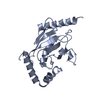
| ||||||||
| 2 | 
| ||||||||
| 3 | 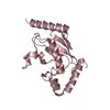
| ||||||||
| Unit cell |
|
-Components
| #1: Protein | Mass: 19059.732 Da / Num. of mol.: 3 Source method: isolated from a genetically manipulated source Source: (gene. exp.) Homo sapiens (human) / Gene: UBE2G2 / Plasmid: pET-cMBP-Gateway / Species (production host): Escherichia coli / Production host:  Escherichia coli BL21(DE3) (bacteria) / Strain (production host): BL21(DE3) / References: UniProt: P60604, ubiquitin-protein ligase Escherichia coli BL21(DE3) (bacteria) / Strain (production host): BL21(DE3) / References: UniProt: P60604, ubiquitin-protein ligase#2: Water | ChemComp-HOH / | Water Mass: 18.015 Da / Num. of mol.: 23 / Source method: isolated from a natural source / Formula: H2O Mass: 18.015 Da / Num. of mol.: 23 / Source method: isolated from a natural source / Formula: H2O |
|---|
-Experimental details
-Experiment
| Experiment | Method: X-RAY DIFFRACTION / Number of used crystals: 1 |
|---|
- Sample preparation
| Crystal | Density Matthews: 3.83 Å3/Da / Density % sol: 67.91 % |
|---|---|
| Crystal grow | Temperature: 293 K / Method: vapor diffusion, hanging drop / pH: 8.1 Details: 12% Glycerol, 0.1M Tris, 1.45M Ammonium Sulfate, pH 8.1, VAPOR DIFFUSION, HANGING DROP, temperature 293K |
-Data collection
| Diffraction | Mean temperature: 100 K |
|---|---|
| Diffraction source | Source: SYNCHROTRON / Site: SPring-8  / Beamline: BL26B1 / Wavelength: 1 Å / Beamline: BL26B1 / Wavelength: 1 Å |
| Detector | Type: RIGAKU JUPITER 210 / Detector: CCD / Date: Dec 17, 2004 / Details: two dimensional focusing mirror |
| Radiation | Monochromator: Si double crystal / Protocol: SINGLE WAVELENGTH / Monochromatic (M) / Laue (L): M / Scattering type: x-ray |
| Radiation wavelength | Wavelength: 1 Å / Relative weight: 1 |
| Reflection | Resolution: 2.56→50 Å / Num. obs: 28705 / % possible obs: 97.5 % / Observed criterion σ(I): -3 / Redundancy: 4.11 % / Biso Wilson estimate: 45.8 Å2 / Rsym value: 0.055 / Net I/σ(I): 22.2 |
| Reflection shell | Resolution: 2.56→2.65 Å / Redundancy: 3.7 % / Mean I/σ(I) obs: 4.2 / Num. unique all: 2445 / Rsym value: 0.293 / % possible all: 84.6 |
- Processing
| Software |
| ||||||||||||||||||||||||||||||||||||
|---|---|---|---|---|---|---|---|---|---|---|---|---|---|---|---|---|---|---|---|---|---|---|---|---|---|---|---|---|---|---|---|---|---|---|---|---|---|
| Refinement | Method to determine structure: MOLECULAR REPLACEMENT Starting model: PDB ENTRY 2UCZ Resolution: 2.56→49.43 Å / Rfactor Rfree error: 0.005 / Data cutoff high absF: 1671970.03 / Data cutoff low absF: 0 / Isotropic thermal model: RESTRAINED / Cross valid method: THROUGHOUT / σ(F): 0 / Stereochemistry target values: MAXIMUM LIKELIHOOD
| ||||||||||||||||||||||||||||||||||||
| Solvent computation | Solvent model: FLAT MODEL / Bsol: 55.643 Å2 / ksol: 0.355828 e/Å3 | ||||||||||||||||||||||||||||||||||||
| Displacement parameters | Biso mean: 75.7 Å2
| ||||||||||||||||||||||||||||||||||||
| Refine analyze |
| ||||||||||||||||||||||||||||||||||||
| Refinement step | Cycle: LAST / Resolution: 2.56→49.43 Å
| ||||||||||||||||||||||||||||||||||||
| Refine LS restraints |
| ||||||||||||||||||||||||||||||||||||
| LS refinement shell | Resolution: 2.56→2.72 Å / Rfactor Rfree error: 0.02 / Total num. of bins used: 6
| ||||||||||||||||||||||||||||||||||||
| Xplor file |
|
