 Movie
Movie Controller

[English] 日本語
 Yorodumi
- PDB-2csw: Solution structure of the FHA domain of human ubiquitin ligase pr... -
+ Open data

- Basic information
| Entry | Database: PDB / ID: 2csw | ||||||
|---|---|---|---|---|---|---|---|
| Title | Solution structure of the FHA domain of human ubiquitin ligase protein RNF8 | ||||||
 Components Components | Ubiquitin ligase protein RNF8 | ||||||
 Keywords Keywords | LIGASE / 11-stranded beta sandwich / RING finger protein 8 / Structural Genomics / NPPSFA / National Project on Protein Structural and Functional Analyses / RIKEN Structural Genomics/Proteomics Initiative / RSGI | ||||||
| Function / homology |  Function and homology information Function and homology informationsperm DNA condensation / isotype switching / DNA repair-dependent chromatin remodeling / response to ionizing radiation / negative regulation of transcription elongation by RNA polymerase II / protein K63-linked ubiquitination / protein K48-linked ubiquitination / protein autoubiquitination / interstrand cross-link repair / ubiquitin ligase complex ...sperm DNA condensation / isotype switching / DNA repair-dependent chromatin remodeling / response to ionizing radiation / negative regulation of transcription elongation by RNA polymerase II / protein K63-linked ubiquitination / protein K48-linked ubiquitination / protein autoubiquitination / interstrand cross-link repair / ubiquitin ligase complex / epigenetic regulation of gene expression / signal transduction in response to DNA damage / positive regulation of DNA repair / ubiquitin binding / Nonhomologous End-Joining (NHEJ) / RING-type E3 ubiquitin transferase / G2/M DNA damage checkpoint / double-strand break repair via nonhomologous end joining / ubiquitin-protein transferase activity / ubiquitin protein ligase activity / double-strand break repair / Recruitment and ATM-mediated phosphorylation of repair and signaling proteins at DNA double strand breaks / site of double-strand break / Processing of DNA double-strand break ends / midbody / histone binding / ubiquitin-dependent protein catabolic process / chromosome, telomeric region / cell cycle / cell division / DNA damage response / chromatin binding / ubiquitin protein ligase binding / protein homodimerization activity / zinc ion binding / nucleoplasm / identical protein binding / nucleus / cytosolSimilarity search - Function | ||||||
| Biological species |  Homo sapiens (human) Homo sapiens (human) | ||||||
| Method | SOLUTION NMR / torsion angle dynamics | ||||||
 Authors Authors | Hayashi, F. / Nagashima, T. / Yokoyama, S. / RIKEN Structural Genomics/Proteomics Initiative (RSGI) | ||||||
 Citation Citation | Journal: To be Published Title: Solution structure of the FHA domain of human ubiquitin ligase protein RNF8 Authors: Hayashi, F. / Nagashima, T. / Yokoyama, S. | ||||||
| History |
|
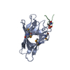
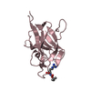
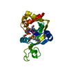
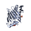
- Structure visualization
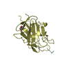
| Structure viewer | Molecule: MolmilJmol/JSmol |
|---|
- Downloads & links
-Download
| PDBx/mmCIF format | 2csw.cif.gz | 851.9 KB | Display | PDBx/mmCIF format |
|---|---|---|---|---|
| PDB format | pdb2csw.ent.gz | 741.2 KB | Display | PDB format |
| PDBx/mmJSON format | 2csw.json.gz | Tree view | PDBx/mmJSON format | |
| Others |  Other downloads Other downloads |
-Validation report
| Arichive directory | https://data.pdbj.org/pub/pdb/validation_reports/cs/2cswftp://data.pdbj.org/pub/pdb/validation_reports/cs/2csw | HTTPS FTP |
|---|
-Related structure data
| Similar structure data | |
|---|---|
| Other databases |


-Links
 PDBj
PDBj


- Assembly
| Deposited unit | 
| |||||||||
|---|---|---|---|---|---|---|---|---|---|---|
| 1 |
| |||||||||
| NMR ensembles |
|
-Components
| #1: Protein | / RING finger protein 8 Mass: 16056.050 Da / Num. of mol.: 1 / Fragment: FHA domain Source method: isolated from a genetically manipulated source Source: (gene. exp.) Homo sapiens (human) / Description: Cell-free protein synthesis / Gene: RNF8 / Plasmid: P040322-86References: UniProt: O76064, Ligases; Forming carbon-nitrogen bonds; Acid-amino-acid ligases (peptide synthases) |
|---|
-Experimental details
-Experiment
| Experiment | Method: SOLUTION NMR | ||||||||||||
|---|---|---|---|---|---|---|---|---|---|---|---|---|---|
| NMR experiment |
| ||||||||||||
| NMR details | Text: The structure was determined using triple-resonance NMR spectroscopy. |
- Sample preparation
| Details | Contents: 1.1mM 13C,15N-labeled protein; 20mM d-TrisHCl; 100mM NaCl; 1mM d-DTT; 0.02% NaN3 Solvent system: 90% H2O/10% D2O |
|---|---|
| Sample conditions | Ionic strength: 120mM / pH: 7.0 / Pressure: ambient / Temperature: 298 K |
-NMR measurement
| NMR spectrometer |
|
|---|
- Processing
| NMR software |
| ||||||||||||||||||||||||||||
|---|---|---|---|---|---|---|---|---|---|---|---|---|---|---|---|---|---|---|---|---|---|---|---|---|---|---|---|---|---|
| Refinement | Method: torsion angle dynamics / Software ordinal: 1 | ||||||||||||||||||||||||||||
| NMR representative | Selection criteria: lowest energy | ||||||||||||||||||||||||||||
| NMR ensemble | Conformer selection criteria: target function, structures with the lowest energy, structures with the least restraint violations Conformers calculated total number: 100 / Conformers submitted total number: 20 |
