 Movie
Movie Controller

[English] 日本語
 Yorodumi
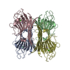
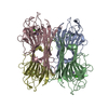
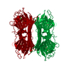
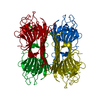
- PDB-2cna: THE COVALENT AND THREE-DIMENSIONAL STRUCTURE OF CONCANAVALIN A, I... -
+ Open data

- Basic information
| Entry | Database: PDB / ID: 2cna | ||||||
|---|---|---|---|---|---|---|---|
| Title | THE COVALENT AND THREE-DIMENSIONAL STRUCTURE OF CONCANAVALIN A, IV.ATOMIC COORDINATES,HYDROGEN BONDING,AND QUATERNARY STRUCTURE | ||||||
 Components Components | CONCANAVALIN A | ||||||
 Keywords Keywords | LECTIN (AGGLUTININ) | ||||||
| Function / homology |  Function and homology information Function and homology informationregulation of defense response to virus / mannose binding / defense response / metal ion bindingSimilarity search - Function | ||||||
| Biological species |   Canavalia ensiformis (jack bean) Canavalia ensiformis (jack bean) | ||||||
| Method | X-RAY DIFFRACTION / Resolution: 2 Å | ||||||
 Authors Authors | Reekejunior, G.N. / Becker, J.W. / Edelman, G.M. | ||||||
 Citation Citation | Journal: J.Biol.Chem. / Year: 1975 Title: The covalent and three-dimensional structure of concanavalin A. IV. Atomic coordinates, hydrogen bonding, and quaternary structure. Authors: Reeke Jr., G.N. / Becker, J.W. / Edelman, G.M. #1: Journal: J.Biol.Chem. / Year: 1975Title: The Covalent and Three-Dimensional Structure of Concanavalin A, I.Amino Acid Sequence of Cyanogen Bromide Fragments F1 and F2 Authors: Wang, J.L. / Cunningham, B.A. / Waxdal, M.J. / Edelman, G.M. #2: Journal: J.Biol.Chem. / Year: 1975Title: The Covalent and Three-Dimensional Structure of Concanavalin A, II.Amino Acid Sequence of Cyanogen Bromide Fragment F3 Authors: Cunningham, B.A. / Wang, J.L. / Waxdal, M.J. / Edelman, G.M. #3: Journal: J.Biol.Chem. / Year: 1975Title: The Covalent and Three-Dimensional Structure of Concanavalin A, III.Structure of the Monomer and its Interactions with Metals and Saccharides Authors: Becker, J.W. / Reekejunior, G.N. / Wang, J.L. / Cunningham, B.A. / Edelman, G.M. | ||||||
| History |
| ||||||
| Remark 700 | SHEET THE MOLECULE CONTAINS TWO PLEATED SHEET STRUCTURES OF SEVEN ANTIPARALLEL STRANDS EACH. TWO ...SHEET THE MOLECULE CONTAINS TWO PLEATED SHEET STRUCTURES OF SEVEN ANTIPARALLEL STRANDS EACH. TWO STRANDS (5 AND 6) OF THE SHEET DESIGNATED *BK* AND ONE STRAND (6) OF THE SHEET DESIGNATED *FT* CONTAIN RESIDUES FROM DISCONTINUOUS PARTS OF THE AMINO ACID SEQUENCE. IN THE FORMAT SPECIFIED BY THE DATA BANK FOR REPRESENTING SUCH SHEETS ON SHEET CARDS, EACH SHEET IS LISTED TWICE. THE SINGLE *BK* SHEET IS LISTED ONCE AS *BK1* AND ONCE AS *BK2*, AND THE SINGLE *FT* SHEET IS LISTED ONCE AS *FT1* AND ONCE AS *FT2*. THESE REDUNDANT DESCRIPTIONS DO NOT IMPLY THE PRESENCE OF MORE THAN TWO SHEETS IN THE STRUCTURE. STRANDS 1,2,3,4 OF BK1 ARE THE SAME AS STRANDS 1,2,3,4 OF BK2. SIMILARLY, STRANDS 1,2,3, 4,5 OF FT1 ARE THE SAME AS STRANDS 1,2,3,4,5 OF FT2. THE NON-IDENTICAL STRANDS DESCRIBE THE DISJOINT SEGMENTS OF STRANDS 5 AND 6 OF THE *BK* SHEET AND STRAND 6 OF THE *FT* SHEET. IN ORDER TO PRESENT THE PLEATED SHEET STRUCTURE OF CON A IN A MORE EASILY UNDERSTOOD FORMAT, THE FOLLOWING REMARK CARDS CONTAIN A DESCRIPTION OF THE SHEETS LIKE THAT ON THE SHEET CARDS, EXCEPT THAT EACH SHEET IS LISTED ONCE ONLY, AND DISJOINT STRANDS ARE DESIGNATED BY LETTERS, E.G. 5A, 5B ETC. 1 BK 7 THR 73 VAL 79 0 2 BK 7 LYS 59 SER 66 -1 N VAL 75 O ALA 63 3 BK 7 GLY 48 SER 56 -1 N VAL 64 O HIS 51 4 BK 7 VAL 188 LYS 200 -1 N ILE 52 O ALA 193 5A BK 7 SER 108 LYS 116 -1 N GLU 192 O THR 112 5B BK 7 THR 103 ILE 106 -1 N LEU 198 O THR 105 6A BK 7 ASP 124 PHE 130 -1 N SER 113 O LEU 126 6B BK 7 ASN 153 LEU 156 -1 N ILE 106 O LEU 154 7 BK 7 THR 147 GLY 149 -1 N GLU 155 O THR 147 1 FNT 7 LYS 35 TRP 40 0 2 FNT 7 PRO 23 LYS 30 -1 N ALA 38 O ILE 25 3 FNT 7 ILE 4 THR 11 -1 N GLY 26 O GLU 8 4 FNT 7 ASP 208 SER 215 -1 N VAL 7 O PHE 212 5 FNT 7 GLU 87 THR 97 -1 N PHE 213 O GLY 92 6AFNT 7 SER 169 TYR 176 -1 N LEU 93 O ALA 173 6BFNT 7 VAL 179 ILE 181 -1 N VAL 89 O VAL 179 7 FNT 7 ASP 139 GLN 143 -1 N LEU 174 O ILE 141 SEVERAL RESIDUES WITHIN EACH SHEET STRUCTURE HAVE UNUSUAL CONFORMATIONS OR FORM UNUSUAL HYDROGEN BONDS. THESE PECULIARITIES ARE DOCUMENTED IN THE REFERENCES GIVEN. STRAND 6 OF SHEET BK1 HYDROGEN BONDS TO A SYMMETRY-RELATED STRAND IN ANOTHER PROTOMER TO FORM A 12-STRANDED ANTI- PARALLEL SHEET IN THE DIMER. |
- Structure visualization
| Structure viewer | Molecule: MolmilJmol/JSmol |
|---|
- Downloads & links
-Download
| PDBx/mmCIF format | 2cna.cif.gz | 55.9 KB | Display | PDBx/mmCIF format |
|---|---|---|---|---|
| PDB format | pdb2cna.ent.gz | 38.6 KB | Display | PDB format |
| PDBx/mmJSON format | 2cna.json.gz | Tree view | PDBx/mmJSON format | |
| Others |  Other downloads Other downloads |
-Validation report
| Arichive directory | https://data.pdbj.org/pub/pdb/validation_reports/cn/2cnaftp://data.pdbj.org/pub/pdb/validation_reports/cn/2cna | HTTPS FTP |
|---|
-Related structure data
| Similar structure data |
|---|


-Links
 PDBj
PDBj

- Assembly
| Deposited unit | 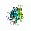
| ||||||||
|---|---|---|---|---|---|---|---|---|---|
| 1 | 
| ||||||||
| Unit cell |
| ||||||||
| Atom site foot note | 1: A001 WEAK DENSITY, INTERPRETATION UNCERTAIN / 2: E008 MN2+ LIGAND; OE1 CLOSE TO D28 OD1 / 3: D010 MN2+ AND CA2+ LIGAND 4: T011 OG JOINED TO E102 OE2 & D208 O -PROB BIFURCATED H-BOND 5: Y012 SIDE CHAIN IS NEAR ANOTHER MOLECULE / 6: N014 CA2+ LIGAND / 7: D016 OD1 WEAK / 8: I017 CG2 WEAK / 9: G018 EXTRA DENSITY AT CA; O - MN2+ SOLVENT BRIDGE / 10: D019 MN2+ AND CA2+ LIGAND; O CLOSE TO P20 O / 11: P020 CG, CD WEAK; O CLOSE TO D19 O / 12: Y022 OH WEAK / 13: P023 CARBONYL WEAK / 14: H024 MN2+ LIGAND; SIDE CHAIN STRONG / 15: I025 CD1 WEAK / 16: D028 CLOSE TO E8 OE1 / 17: K030 SIDE CHAIN DISORDERED BEYOND CD / 18: V032 SIDE CHAIN IS MISSHAPEN; W1 IS MN2+ LIGAND / 19: R033 CB, CG WEAK 20: S034 STRONG DENSITY HARD TO INTERPRET; W2 IS MN2+ LIGAND 21: K039 SIDE CHAIN DISORDERED BEYOND CB / 22: W040 CB WEAK / 23: N041 SIDE CHAIN DISORDERED / 24: M042 CA WEAK / 25: D044 CARBONYL WEAK / 26: K046 SIDE CHAIN DISORDERED BEYOND CD / 27: S056 CARBONYL WEAK / 28: D058 DENSITY VERY STRONG; TETRAMER SALT BRIDGE 29: R060 DENSITY CONFUSING AT GUANIDINIUM; TETRAMER SALT BRIDGE 30: P068 ALMOST SPHERICAL / 31: N069 DENSITY SOMEWHAT SPREAD OUT / 32: A072 DENSITY DISTORTED AT CB / 33: T073 NO DENSITY FOR SIDE CHAIN / 34: S076 CARBONYL WEAK / 35: D080 SIDE CHAIN CONFUSING / 36: N082 SIDE CHAIN WEAK / 37: L085 SIDE CHAIN CONFUSING / 38: P086 CG AND CD WEAK / 39: E087 CG WEAK / 40: V089 SIDE CHAIN DISORDERED / 41: A095 PROBABLY IS CLOSER TO G171 42: G098 STRONG BRIDGE G98 N - Y100 O MAKES INTERP DIFFICULT 43: L099 SIDE CHAIN DENSITY ALMOST ABSENT / 44: T105 SIDE CHAIN DISORDERED / 45: S108 OG POSITION UNCLEAR / 46: K114 CD WEAK; TETRAMER SLAT LINK / 47: K116 C, CD WEAK; TETRAMER SLAT LINK / 48: S117 SIDE CHAIN WEAK / 49: S119 DENSITY CONFUSING; INTERPRETATION UNCERTAIN / 50: T120 DENSITY CONFUSING; INTERPRETATION UNCERTAIN / 51: H121 SIDE CHAIN AND CARBONYL WEAK / 52: Q122 N, CB WEAK; LOCAL DISORDER AT 2-FOLD PARALLEL TO X / 53: T123 SIDE CHAIN CONFUSING / 54: H127 LARGE DENSITY AT SITE OF NEARBY METAL SUBSTITUENTS / 55: M129 SIDE CHAIN DENSITY JOINS H127; SEE H127 / 56: Q132 CB WEAK / 57: K135 CARBONYL CONFUSING / 58: D136 SIDE CHAIN DISORDERED / 59: Q137 CONFORMATION PECULIAR; INTERPRETATION UNCERTAIN / 60: A146 CB WEAK / 61: T147 SIDE CHAIN DENSITY CONFUSING / 62: T148 LARGE DENSITY AT CARBONYL; PROBABLE DISORDER / 63: T150 SIDE CHAIN DENSITY NOT IDENTIFIABLE / 64: D151 N WEAK / 65: L154 N WEAK / 66: V159 CG1, CG2 WEAK / 67: S160 SIDE CHAIN CONFUSING; AMIDE H-BOND BIFURCATED / 68: S161 DENSITY CONFUSING / 69: N162 DENSITY CONFUSING; INTERPRETATION UNCERTAIN / 70: E166 SIDE CHAIN DENSITY AT CARBOXYL ONLY / 71: S168 ORIENTATION OF OG UNCLEAR / 72: S169 OG H-BOND BIFURCATED / 73: Y176 CE2 WEAK; CARBONYL STRONG / 74: A177 CARBONYL WEAK; APPEARS OUT OF PLANE OF P178 / 75: H180 O WEAK / 76: I181 CG2 HAS NO DENSITY / 77: S184 SIDE CHAIN DISORDERED; NEAR ANOTHER MOLECULE / 78: S185 SIDE CHAIN CONFUSING / 79: T187 DENSITY CONFUSING - LARGE SIDE CHAIN PRESENT / 80: S189 OG WEAK / 81: E192 TETRAMER SALT LINKS 82: T194 SIDE CHAIN DISORDERED; JOINS H51; AMIDE H-BOND BIFURC? 83: A196 SIDE CHAIN DISORDERED / 84: L198 SIDE CHAIN SLIGHTLY DISORDERED / 85: K200 DISORDERED BEYOND CD / 86: S201 AMIDE H-BOND IS BIFURCATED / 87: A207 POSSIBLE CIS-PEPTIDE / 88: N216 ORIENTATION OF CARBOXAMIDO GROUP UNCERTAIN / 89: S220 PEPTIDE CONFUSING; AMIDE H-BOND BIFURCATED / 90: I221 SIDE CHAIN WEAK / 91: S223 SIDE CHAIN DISORDERED / 92: G224 CARBONYL WEAK / 93: S225 OG DISORDERED / 94: R228 CARBONYL WEAK; GUANIDINIUM GROUP CONFUSING / 95: L230 SIDE CHAIN VERY WEAK BEYOND CB / 96: L232 SIDE CHAIN CONFUSING / 97: F233 CZ WEAK 98: D235 - N237 APPEAR TO EXIST IN 2 CONFORMATIONS- NEITHER FIT IS VERY GOOD; SIDE CHAIN OBSCURE |
-Components
| #1: Protein | Mass: 25596.299 Da / Num. of mol.: 1 Source method: isolated from a genetically manipulated source Source: (gene. exp.) Canavalia ensiformis (jack bean) / References: UniProt: P02866 |
|---|---|
| #2: Chemical | ChemComp-MN /   Mass: 54.938 Da / Num. of mol.: 1 / Source method: obtained synthetically / Formula: Mn Mass: 54.938 Da / Num. of mol.: 1 / Source method: obtained synthetically / Formula: Mn |
| #3: Chemical | ChemComp-CA /   Mass: 40.078 Da / Num. of mol.: 1 / Source method: obtained synthetically / Formula: Ca Mass: 40.078 Da / Num. of mol.: 1 / Source method: obtained synthetically / Formula: Ca |
| #4: Water | ChemComp-HOH / Water Mass: 18.015 Da / Num. of mol.: 4 / Source method: isolated from a natural source / Formula: H2O Mass: 18.015 Da / Num. of mol.: 4 / Source method: isolated from a natural source / Formula: H2O |
| Compound details | ALL SEQUENCES OF 4 RESIDUES FOR WHICH THE DISTANCE CA(I)-CA (I+3) IS LESS THAN 7 ANGSTROMS ARE ...ALL SEQUENCES OF 4 RESIDUES FOR WHICH THE DISTANCE CA(I)-CA (I+3) IS LESS THAN 7 ANGSTROMS ARE LISTED BELOW IN THE TURN RECORDS. THOSE TURNS WHICH PROBABLY CONTAIN THE HYDROGEN BOND N(I+3)...O(I) ARE DESIGNATED |
-Experimental details
-Experiment
| Experiment | Method: X-RAY DIFFRACTION |
|---|
- Sample preparation
| Crystal | Density Matthews: 2.41 Å3/Da / Density % sol: 49.06 % | ||||||||||||||||||
|---|---|---|---|---|---|---|---|---|---|---|---|---|---|---|---|---|---|---|---|
| Crystal grow | *PLUS pH: 6.8 / Method: unknown / Details: referred to J.Biol.Chem. 250.1513-1524 1975 | ||||||||||||||||||
| Components of the solutions | *PLUS
|
- Processing
| Refinement | Highest resolution: 2 Å | ||||||||||||
|---|---|---|---|---|---|---|---|---|---|---|---|---|---|
| Refinement step | Cycle: LAST / Highest resolution: 2 Å
| ||||||||||||
| Refinement | *PLUS Num. reflection obs: 10221 / Rfactor obs: 0.411 | ||||||||||||
| Solvent computation | *PLUS | ||||||||||||
| Displacement parameters | *PLUS |
