 Movie
Movie Controller


+ Open data

- Basic information
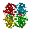
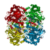
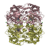
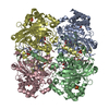
| Entry | Database: PDB / ID: 2cag | ||||||
|---|---|---|---|---|---|---|---|
| Title | CATALASE COMPOUND II | ||||||
 Components Components | CATALASE COMPOUND II | ||||||
 Keywords Keywords | OXIDOREDUCTASE (H2O2 ACCEPTOR) /  PEROXIDASE / IRON / HEME / HYDROGEN PEROXIDE / NADP PEROXIDASE / IRON / HEME / HYDROGEN PEROXIDE / NADP | ||||||
| Function / homology |  Function and homology informationcatalase / catalase activity / hydrogen peroxide catabolic process / response to oxidative stress / heme binding / metal ion binding / cytoplasm Function and homology informationcatalase / catalase activity / hydrogen peroxide catabolic process / response to oxidative stress / heme binding / metal ion binding / cytoplasmSimilarity search - Function | ||||||
| Biological species |  Proteus mirabilis (bacteria) Proteus mirabilis (bacteria) | ||||||
| Method | X-RAY DIFFRACTION / SYNCHROTRON / MOLECULAR REPLACEMENT / Resolution: 2.7 Å | ||||||
 Authors Authors | Gouet, P. / Jouve, H.M. / Hajdu, J. | ||||||
 Citation Citation | Journal: Nat.Struct.Biol. / Year: 1996 Title: Ferryl intermediates of catalase captured by time-resolved Weissenberg crystallography and UV-VIS spectroscopy. Authors: Gouet, P. / Jouve, H.M. / Williams, P.A. / Andersson, I. / Andreoletti, P. / Nussaume, L. / Hajdu, J. #1: Journal: J.Mol.Biol. / Year: 1995Title: Crystal Structure of Proteus Mirabilis Pr Catalase with and without Bound Nadph Authors: Gouet, P. / Jouve, H.M. / Dideberg, O. #2: Journal: Acta Crystallogr.,Sect.B / Year: 1986Title: The Refined Structure of Beef Liver Catalase at 2.5 Angstroms Resolution Authors: Fita, I. / Silva, A.M. / Murthy, M.R.N. / Rossmann, M.G. | ||||||
| History |
|
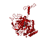
- Structure visualization
| Structure viewer | Molecule: MolmilJmol/JSmol |
|---|
- Downloads & links
-Download
| PDBx/mmCIF format | 2cag.cif.gz | 108 KB | Display | PDBx/mmCIF format |
|---|---|---|---|---|
| PDB format | pdb2cag.ent.gz | 85.9 KB | Display | PDB format |
| PDBx/mmJSON format | 2cag.json.gz | Tree view | PDBx/mmJSON format | |
| Others |  Other downloads Other downloads |
-Validation report
| Arichive directory | https://data.pdbj.org/pub/pdb/validation_reports/ca/2cagftp://data.pdbj.org/pub/pdb/validation_reports/ca/2cag | HTTPS FTP |
|---|
-Related structure data
| Related structure data |  2cae S: Starting model for refinement |
|---|---|
| Similar structure data |


-Links
 PDBj
PDBj
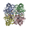
- Assembly
| Deposited unit | 
| ||||||||
|---|---|---|---|---|---|---|---|---|---|
| 1 | 
| ||||||||
| Unit cell |
| ||||||||
| Components on special symmetry positions |
|
-Components
| #1: Protein | Mass: 55726.348 Da / Num. of mol.: 1 / Source method: isolated from a natural source Details: COMPOUND II WAS OBTAINED BY REDUCING A CRYSTAL CONTAINING THE PROTEIN IN THE COMPOUND I STATE WITH DITHIOTHREITOL Source: (natural) Proteus mirabilis (bacteria) / Organ: LIVER / Variant: PEROXIDE RESISTANT MUTANT / References: UniProt: P42321, catalase |
|---|---|
| #2: Chemical | ChemComp-HEM / Heme B  Mass: 616.487 Da / Num. of mol.: 1 / Source method: obtained synthetically / Formula: C34H32FeN4O4 Mass: 616.487 Da / Num. of mol.: 1 / Source method: obtained synthetically / Formula: C34H32FeN4O4 |
| #3: Water | ChemComp-HOH / Water Mass: 18.015 Da / Num. of mol.: 70 / Source method: isolated from a natural source / Formula: H2O Mass: 18.015 Da / Num. of mol.: 70 / Source method: isolated from a natural source / Formula: H2O |
| Compound details | OXYGEN OF THE PROXIMAL TYROSINE 337 IS DEPROTONAT |
-Experimental details
-Experiment
| Experiment | Method: X-RAY DIFFRACTION / Number of used crystals: 1 |
|---|
- Sample preparation
| Crystal | Density Matthews: 4.05 Å3/Da / Density % sol: 63 % | ||||||||||||||||||||||||||||||||||||||||||
|---|---|---|---|---|---|---|---|---|---|---|---|---|---|---|---|---|---|---|---|---|---|---|---|---|---|---|---|---|---|---|---|---|---|---|---|---|---|---|---|---|---|---|---|
| Crystal grow | *PLUS Temperature: 4-5 ℃ / pH: 7.5 / Method: vapor diffusionDetails: drop solution was mixed with an equal volume of reservoir solution | ||||||||||||||||||||||||||||||||||||||||||
| Components of the solutions | *PLUS
|
-Data collection
| Diffraction | Mean temperature: 290 K |
|---|---|
| Diffraction source | Source: SYNCHROTRON / Site: Photon Factory  / Beamline: BL-6A / Wavelength: 1 / Beamline: BL-6A / Wavelength: 1 |
| Detector | Detector: FILM / Date: 1994 |
| Radiation | Monochromatic (M) / Laue (L): M / Scattering type: x-ray |
| Radiation wavelength | Wavelength: 1 Å / Relative weight: 1 |
| Reflection | Resolution: 2.7→30 Å / Num. obs: 19251 / % possible obs: 73 % / Observed criterion σ(I): 0 / Redundancy: 2.1 % / Rmerge(I) obs: 0.076 |
| Reflection shell | Resolution: 2.7→2.8 Å / Rmerge(I) obs: 0.42 / % possible all: 75 |
| Reflection | *PLUS Num. measured all: 39971 |
| Reflection shell | *PLUS % possible obs: 75 % |
- Processing
| Software |
| ||||||||||||||||||||||||||||||||||||||||||||||||||||||||||||
|---|---|---|---|---|---|---|---|---|---|---|---|---|---|---|---|---|---|---|---|---|---|---|---|---|---|---|---|---|---|---|---|---|---|---|---|---|---|---|---|---|---|---|---|---|---|---|---|---|---|---|---|---|---|---|---|---|---|---|---|---|---|
| Refinement | Method to determine structure: MOLECULAR REPLACEMENT Starting model: 2CAE 2cae Resolution: 2.7→15 Å / Cross valid method: FREE R / σ(F): 2 Details: COORDINATES FOR RESIDUES 1 - 3 AND 479 - 484 ARE NOT INCLUDED. COORDINATES FOR SIDE CHAINS OF RESIDUES 204, 395, 451 AND 72, 450, 473 ARE NOT OBSERVED BEYOND CARBON CB AND CG RESPECTIVELY ...Details: COORDINATES FOR RESIDUES 1 - 3 AND 479 - 484 ARE NOT INCLUDED. COORDINATES FOR SIDE CHAINS OF RESIDUES 204, 395, 451 AND 72, 450, 473 ARE NOT OBSERVED BEYOND CARBON CB AND CG RESPECTIVELY AND MODELED WITH AN OCCUPANCY OF 0.00 AND A BFACTOR OF 99.99.
| ||||||||||||||||||||||||||||||||||||||||||||||||||||||||||||
| Displacement parameters | Biso mean: 29 Å2 | ||||||||||||||||||||||||||||||||||||||||||||||||||||||||||||
| Refinement step | Cycle: LAST / Resolution: 2.7→15 Å
| ||||||||||||||||||||||||||||||||||||||||||||||||||||||||||||
| Refine LS restraints |
| ||||||||||||||||||||||||||||||||||||||||||||||||||||||||||||
| Xplor file |
| ||||||||||||||||||||||||||||||||||||||||||||||||||||||||||||
| Software | *PLUS Name: X-PLOR / Classification: refinement | ||||||||||||||||||||||||||||||||||||||||||||||||||||||||||||
| Refinement | *PLUS | ||||||||||||||||||||||||||||||||||||||||||||||||||||||||||||
| Solvent computation | *PLUS | ||||||||||||||||||||||||||||||||||||||||||||||||||||||||||||
| Displacement parameters | *PLUS | ||||||||||||||||||||||||||||||||||||||||||||||||||||||||||||
| Refine LS restraints | *PLUS
|
