 Movie
Movie Controller


+ Open data

- Basic information
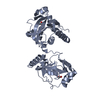
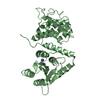
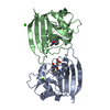
| Entry | Database: PDB / ID: 2ca6 | ||||||
|---|---|---|---|---|---|---|---|
| Title | MIRAS structure determination from hemihedrally twinned crystals | ||||||
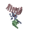
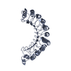
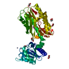
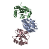
 Components Components | RAN GTPASE-ACTIVATING PROTEIN 1 | ||||||
 Keywords Keywords | SIGNALING REGULATOR / GAP / GTPASE ACTIVATION /  GTPASE-ACTIVATING PROTEIN / HEMIHEDRAL TWINNING / LEUCINE-RICH REPEAT PROTEIN / LRR / MEROHEDRAL TWINNING / MEROHEDRY / RANGAP / RNA1P / SIGNALING PROTEIN / SIGNALING ACTIVATOR / NUCLEAR TRANSPORT GTPASE-ACTIVATING PROTEIN / HEMIHEDRAL TWINNING / LEUCINE-RICH REPEAT PROTEIN / LRR / MEROHEDRAL TWINNING / MEROHEDRY / RANGAP / RNA1P / SIGNALING PROTEIN / SIGNALING ACTIVATOR / NUCLEAR TRANSPORT | ||||||
| Function / homology |  Function and homology information Function and homology informationSUMOylation of nuclear envelope proteins / Postmitotic nuclear pore complex (NPC) reformation / nucleocytoplasmic transport / small GTPase-mediated signal transduction / nuclear periphery / GTPase activator activity / positive regulation of protein export from nucleus / small GTPase binding / perinuclear region of cytoplasm / nucleus ...SUMOylation of nuclear envelope proteins / Postmitotic nuclear pore complex (NPC) reformation / nucleocytoplasmic transport / small GTPase-mediated signal transduction / nuclear periphery / GTPase activator activity / positive regulation of protein export from nucleus / small GTPase binding / perinuclear region of cytoplasm / nucleus / cytosol / cytoplasmSimilarity search - Function | ||||||
| Biological species |  SCHIZOSACCHAROMYCES POMBE (fission yeast) SCHIZOSACCHAROMYCES POMBE (fission yeast) | ||||||
| Method | X-RAY DIFFRACTION / SYNCHROTRON / MIRAS / Resolution: 2.2 Å | ||||||
 Authors Authors | Hillig, R.C. / Renault, L. | ||||||
 Citation Citation | Journal: Acta Crystallogr.,Sect.D / Year: 2006 Title: Detecting and Overcoming Hemihedral Twinning During the Mir Structure Determination of RNA1P. Authors: Hillig, R.C. / Renault, L. #1: Journal: Mol.Cell / Year: 1999Title: The Crystal Structure of RNA1P: A New Fold for a Gtpase-Activating Protein Authors: Hillig, R.C. / Renault, L. / Vetter, I.R. / Drell, T. / Wittinghofer, A. / Becker, J. #2: Journal: Nature / Year: 2002Title: Rangap Mediates GTP Hydrolysis without an Arginine Finger Authors: Seewald, M.J. / Korner, C. / Wittinghofer, A. / Vetter, I.R. | ||||||
| History |
|
- Structure visualization
| Structure viewer | Molecule: MolmilJmol/JSmol |
|---|
- Downloads & links
-Download
| PDBx/mmCIF format | 2ca6.cif.gz | 145.6 KB | Display | PDBx/mmCIF format |
|---|---|---|---|---|
| PDB format | pdb2ca6.ent.gz | 121.6 KB | Display | PDB format |
| PDBx/mmJSON format | 2ca6.json.gz | Tree view | PDBx/mmJSON format | |
| Others |  Other downloads Other downloads |
-Validation report
| Arichive directory | https://data.pdbj.org/pub/pdb/validation_reports/ca/2ca6ftp://data.pdbj.org/pub/pdb/validation_reports/ca/2ca6 | HTTPS FTP |
|---|
-Related structure data
| Related structure data | |
|---|---|
| Similar structure data |


-Links
 PDBj
PDBj- Assembly
| Deposited unit | 
| ||||||||
|---|---|---|---|---|---|---|---|---|---|
| 1 | 
| ||||||||
| 2 | 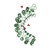
| ||||||||
| Unit cell |
| ||||||||
| Noncrystallographic symmetry (NCS) | NCS oper: (Code: given Matrix: (-0.99729, 0.07335, 0.00611), Vector : |
-Components
| #1: Protein | Mass: 43272.652 Da / Num. of mol.: 2 Source method: isolated from a genetically manipulated source Details: C-TERMINI OF BOTH COPIES OF RNA1P (RESIDUES 345-386) ARE DISORDERED AND WERE OMITTED FROM THE MODEL Source: (gene. exp.) SCHIZOSACCHAROMYCES POMBE (fission yeast)Production host:  ESCHERICHIA COLI (E. coli) / Strain (production host): BL21 / References: UniProt: P41391 ESCHERICHIA COLI (E. coli) / Strain (production host): BL21 / References: UniProt: P41391#2: Chemical | ChemComp-SO4 / Sulfate  Mass: 96.063 Da / Num. of mol.: 4 / Source method: obtained synthetically / Formula: SO4 Mass: 96.063 Da / Num. of mol.: 4 / Source method: obtained synthetically / Formula: SO4#3: Water | ChemComp-HOH / | Water Mass: 18.015 Da / Num. of mol.: 273 / Source method: isolated from a natural source / Formula: H2O Mass: 18.015 Da / Num. of mol.: 273 / Source method: isolated from a natural source / Formula: H2OCompound details | GTPASE ACTIVATOR FOR THE NUCLEAR RAS-RELATED REGULATORY PROTEIN SPI1 (RAN), CONVERTING IT TO THE ...GTPASE ACTIVATOR FOR THE NUCLEAR RAS-RELATED REGULATORY | Sequence details | THE CONFLICT SHOWN IN THE SEQADV RECORDS BELOW SER2ALA IS A CLONING ARTEFACT DUE TO EXPRESSION ...THE CONFLICT SHOWN IN THE SEQADV RECORDS BELOW SER2ALA IS A CLONING ARTEFACT DUE TO EXPRESSION | |
|---|
-Experimental details
-Experiment
| Experiment | Method: X-RAY DIFFRACTION / Number of used crystals: 1 |
|---|
- Sample preparation
| Crystal | Density Matthews: 2.48 Å3/Da / Density % sol: 55.4 % |
|---|---|
| Crystal grow | pH: 8.5 Details: 2 MICROLITER PROTEIN (25MG/ML IN 20MM TRIS-HCL PH7.5, 2MMDTE) AND 2 MICROLITER RESERVOIR (24% PEG2000MME, 100MM TRIS PH8.5, 200MM LI2SO4, 20MM MGCL2)., pH 8.50 |
-Data collection
| Diffraction | Mean temperature: 100 K |
|---|---|
| Diffraction source | Source: SYNCHROTRON / Site: EMBL/DESY, HAMBURG  / Beamline: BW7A / Wavelength: 1.0093 / Beamline: BW7A / Wavelength: 1.0093 |
| Detector | Type: MARRESEARCH / Detector: IMAGE PLATE / Date: Oct 2, 1996 |
| Radiation | Protocol: SINGLE WAVELENGTH / Monochromatic (M) / Laue (L): M / Scattering type: x-ray |
| Radiation wavelength | Wavelength: 1.0093 Å / Relative weight: 1 |
| Reflection | Resolution: 2.2→31 Å / Num. obs: 42106 / % possible obs: 97.3 % / Observed criterion σ(I): 0 / Redundancy: 3.8 % / Biso Wilson estimate: 14.4 Å2 / Rsym value: 0.14 / Net I/σ(I): 8 |
| Reflection shell | Resolution: 2.2→2.25 Å / Redundancy: 2 % / Mean I/σ(I) obs: 2.1 / Rsym value: 0.37 / % possible all: 87.4 |
- Processing
| Software |
| ||||||||||||||||||||||||||||||||||||||||||||||||||||||||||||||||||||||||||||||||
|---|---|---|---|---|---|---|---|---|---|---|---|---|---|---|---|---|---|---|---|---|---|---|---|---|---|---|---|---|---|---|---|---|---|---|---|---|---|---|---|---|---|---|---|---|---|---|---|---|---|---|---|---|---|---|---|---|---|---|---|---|---|---|---|---|---|---|---|---|---|---|---|---|---|---|---|---|---|---|---|---|---|
| Refinement | Method to determine structure: MIRAS / Resolution: 2.2→31 Å / Cross valid method: THROUGHOUT / σ(F): 0 Details: THE DATA SET SHOWS HEMIHEDRAL TWINNING WITH A HIGH TWIN FRACTION OF 0.39. THE MODEL WAS REFINED AGAINST THIS ORIGINAL DATA SET, I.E. REFLECTIONS NOT TWIN CORRECTED, BY USING CNX IN TWIN MODE. ...Details: THE DATA SET SHOWS HEMIHEDRAL TWINNING WITH A HIGH TWIN FRACTION OF 0.39. THE MODEL WAS REFINED AGAINST THIS ORIGINAL DATA SET, I.E. REFLECTIONS NOT TWIN CORRECTED, BY USING CNX IN TWIN MODE. ALL R FACTORS ARE SO CALLED TWINNED R FACTORS, CALCULATED FROM THE DIFFERENCES BETWEEN THE ORIGINAL TWINNED REFLECTIONS AND THE ARTIFICIALLY TWINNED REFLECTIONS CALCULATED FROM THE MODEL.
| ||||||||||||||||||||||||||||||||||||||||||||||||||||||||||||||||||||||||||||||||
| Solvent computation | Solvent model: MASK AROUND THE MOLECULE, DETERMINED AUTOMATICALLY BY CNX Bsol: 38.2404 Å2 / ksol: 0.352164 e/Å3 | ||||||||||||||||||||||||||||||||||||||||||||||||||||||||||||||||||||||||||||||||
| Displacement parameters | Biso mean: 22.7 Å2
| ||||||||||||||||||||||||||||||||||||||||||||||||||||||||||||||||||||||||||||||||
| Refine analyze |
| ||||||||||||||||||||||||||||||||||||||||||||||||||||||||||||||||||||||||||||||||
| Refinement step | Cycle: LAST / Resolution: 2.2→31 Å
| ||||||||||||||||||||||||||||||||||||||||||||||||||||||||||||||||||||||||||||||||
| Refine LS restraints |
| ||||||||||||||||||||||||||||||||||||||||||||||||||||||||||||||||||||||||||||||||
| LS refinement shell | Resolution: 2.2→2.24 Å / Total num. of bins used: 20 /
| ||||||||||||||||||||||||||||||||||||||||||||||||||||||||||||||||||||||||||||||||
| Xplor file | Serial no: 1 / Param file: PROTEIN_REP.PARAM / Topol file: ION.TOP |
