 Movie
Movie Controller

[English] 日本語
 Yorodumi
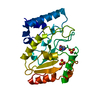
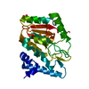
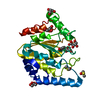
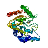
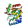
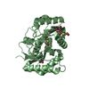
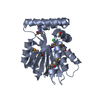
- PDB-2c56: A comparative study of uracil DNA glycosylases from human and her... -
+ Open data

- Basic information
| Entry | Database: PDB / ID: 2c56 | |||||||||
|---|---|---|---|---|---|---|---|---|---|---|
| Title | A comparative study of uracil DNA glycosylases from human and herpes simplex virus type 1 | |||||||||
 Components Components | URACIL DNA GLYCOSYLASE | |||||||||
 Keywords Keywords |  HYDROLASE / URACIL DNA GLYCOSYLASE / DNA REPAIR / DNA DAMAGE / GLYCOSIDASE HYDROLASE / URACIL DNA GLYCOSYLASE / DNA REPAIR / DNA DAMAGE / GLYCOSIDASE | |||||||||
| Function / homology |  Function and homology informationuracil-DNA glycosylase / uracil DNA N-glycosylase activity / base-excision repair / host cell nucleus Function and homology informationuracil-DNA glycosylase / uracil DNA N-glycosylase activity / base-excision repair / host cell nucleusSimilarity search - Function | |||||||||
| Biological species |   HUMAN HERPESVIRUS 1 (Herpes simplex virus type 1) HUMAN HERPESVIRUS 1 (Herpes simplex virus type 1) | |||||||||
| Method | X-RAY DIFFRACTION / SYNCHROTRON / MOLECULAR REPLACEMENT / Resolution: 2.1 Å | |||||||||
 Authors Authors | Krusong, K. / Carpenter, E.P. / Bellamy, S.R.W. / Savva, R. / Baldwin, G.S. | |||||||||
 Citation Citation | Journal: J.Biol.Chem. / Year: 2006 Title: A Comparative Study of Uracil-DNA Glycosylases from Human and Herpes Simplex Virus Type 1. Authors: Krusong, K. / Carpenter, E.P. / Bellamy, S.R.W. / Savva, R. / Baldwin, G.S. | |||||||||
| History |
|
- Structure visualization
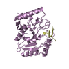
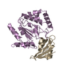
| Structure viewer | Molecule: MolmilJmol/JSmol |
|---|
- Downloads & links
-Download
| PDBx/mmCIF format | 2c56.cif.gz | 67.4 KB | Display | PDBx/mmCIF format |
|---|---|---|---|---|
| PDB format | pdb2c56.ent.gz | 47.1 KB | Display | PDB format |
| PDBx/mmJSON format | 2c56.json.gz | Tree view | PDBx/mmJSON format | |
| Others |  Other downloads Other downloads |
-Validation report
| Arichive directory | https://data.pdbj.org/pub/pdb/validation_reports/c5/2c56ftp://data.pdbj.org/pub/pdb/validation_reports/c5/2c56 | HTTPS FTP |
|---|
-Related structure data
| Related structure data |  2c53C 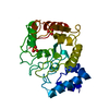 1udgS S: Starting model for refinement C: citing same article ( |
|---|---|
| Similar structure data |


-Links
 PDBj
PDBj- Assembly
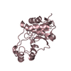
| Deposited unit | 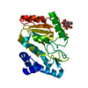
| ||||||||
|---|---|---|---|---|---|---|---|---|---|
| 1 |
| ||||||||
| Unit cell |
|
-Components
| #1: Protein | Mass: 27341.418 Da / Num. of mol.: 1 / Mutation: YES Source method: isolated from a genetically manipulated source Source: (gene. exp.) HUMAN HERPESVIRUS 1 (Herpes simplex virus type 1)Plasmid: D88N/H20N / Production host:  ESCHERICHIA COLI (E. coli) / Strain (production host): BL21 / References: UniProt: P10186, uridine nucleosidase ESCHERICHIA COLI (E. coli) / Strain (production host): BL21 / References: UniProt: P10186, uridine nucleosidase |
|---|---|
| #2: Polysaccharide | beta-D-fructofuranose-(2-1)-alpha-D-glucopyranose / sucrose /   , Oligosaccharide / Class: Nutrient / Mass: 342.297 Da / Num. of mol.: 1 , Oligosaccharide / Class: Nutrient / Mass: 342.297 Da / Num. of mol.: 1Source method: isolated from a genetically manipulated source Details: oligosaccharide with reducing-end-to-reducing-end glycosidic bond References: sucrose |
| #3: Water | ChemComp-HOH / Water Mass: 18.015 Da / Num. of mol.: 259 / Source method: isolated from a natural source / Formula: H2O Mass: 18.015 Da / Num. of mol.: 259 / Source method: isolated from a natural source / Formula: H2O |
| Compound details | CUTS URACIL RESIDUES FROM THE DNA. ENGINEERED RESIDUE IN CHAIN A, ASP 178 TO ASN ENGINEERED RESIDUE ...CUTS URACIL RESIDUES FROM THE DNA. ENGINEERED |
-Experimental details
-Experiment
| Experiment | Method: X-RAY DIFFRACTION |
|---|
- Sample preparation
| Crystal | Density Matthews: 1.89 Å3/Da / Density % sol: 34.51 % |
|---|
-Data collection
| Diffraction | Mean temperature: 100 K |
|---|---|
| Diffraction source | Source: SYNCHROTRON / Site: SRS  / Beamline: PX9.6 / Wavelength: 0.87 / Beamline: PX9.6 / Wavelength: 0.87 |
| Detector | Type: ADSC CCD / Detector: CCD |
| Radiation | Protocol: SINGLE WAVELENGTH / Monochromatic (M) / Laue (L): M / Scattering type: x-ray |
| Radiation wavelength | Wavelength: 0.87 Å / Relative weight: 1 |
| Reflection | Resolution: 2.1→30 Å / Num. obs: 12927 / % possible obs: 100 % / Observed criterion σ(I): -3 / Redundancy: 3.7 % / Biso Wilson estimate: 3.5 Å2 / Rmerge(I) obs: 0.08 / Net I/σ(I): 12.56 |
| Reflection shell | Resolution: 2.1→2.18 Å / Redundancy: 3.7 % / Rmerge(I) obs: 0.22 / Mean I/σ(I) obs: 5.26 / % possible all: 100 |
- Processing
| Software |
| ||||||||||||||||||||||||||||||||||||||||||||||||||||||||||||||||||||||||||||||||
|---|---|---|---|---|---|---|---|---|---|---|---|---|---|---|---|---|---|---|---|---|---|---|---|---|---|---|---|---|---|---|---|---|---|---|---|---|---|---|---|---|---|---|---|---|---|---|---|---|---|---|---|---|---|---|---|---|---|---|---|---|---|---|---|---|---|---|---|---|---|---|---|---|---|---|---|---|---|---|---|---|---|
| Refinement | Method to determine structure: MOLECULAR REPLACEMENT Starting model: PDB ENTRY 1UDG Resolution: 2.1→29.59 Å / Rfactor Rfree error: 0.008 / Data cutoff high absF: 1105069.29 / Isotropic thermal model: RESTRAINED / Cross valid method: THROUGHOUT / σ(F): 0 Details: RESIDUES 1 - 16 IN THIS MOLECULE DO NOT APPEAR IN THE ELECTRON DENSITY AND ARE PRESUMED TO BE DISORDERED.
| ||||||||||||||||||||||||||||||||||||||||||||||||||||||||||||||||||||||||||||||||
| Solvent computation | Solvent model: CNS BULK SOLVENT MODEL USED / Bsol: 52.0358 Å2 / ksol: 0.335261 e/Å3 | ||||||||||||||||||||||||||||||||||||||||||||||||||||||||||||||||||||||||||||||||
| Displacement parameters | Biso mean: 14.71 Å2
| ||||||||||||||||||||||||||||||||||||||||||||||||||||||||||||||||||||||||||||||||
| Refine analyze |
| ||||||||||||||||||||||||||||||||||||||||||||||||||||||||||||||||||||||||||||||||
| Refinement step | Cycle: LAST / Resolution: 2.1→29.59 Å
| ||||||||||||||||||||||||||||||||||||||||||||||||||||||||||||||||||||||||||||||||
| Refine LS restraints |
| ||||||||||||||||||||||||||||||||||||||||||||||||||||||||||||||||||||||||||||||||
| LS refinement shell | Resolution: 2.1→2.23 Å / Rfactor Rfree error: 0.02 / Total num. of bins used: 6
|
