 Movie
Movie Controller


+ Open data

- Basic information
| Entry | Database: PDB / ID: 2buf | |||||||||
|---|---|---|---|---|---|---|---|---|---|---|
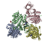
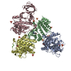
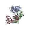
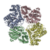
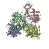
| Title | Arginine Feed-Back Inhibitable Acetylglutamate Kinase | |||||||||
 Components Components | ACETYLGLUTAMATE KINASE | |||||||||
 Keywords Keywords | TRANSFERASE / ACETYGLUTAMATE KINASE / ACETYLGLUTAMATE / ADP / ARGININE BIOSYNTHESIS / FEED-BACK INHIBITION / HEXAMER / ARGININE INHIBITION / ALLOSTERIC MECHANISM / FEEDBACK CONTROL / AMINO ACID KINASE FAMILY | |||||||||
| Function / homology |  Function and homology informationacetylglutamate kinase / acetylglutamate kinase activity / arginine biosynthetic process via ornithine / arginine biosynthetic process / arginine binding / ATP binding / cytoplasm Function and homology informationacetylglutamate kinase / acetylglutamate kinase activity / arginine biosynthetic process via ornithine / arginine biosynthetic process / arginine binding / ATP binding / cytoplasmSimilarity search - Function | |||||||||
| Biological species |   PSEUDOMONAS AERUGINOSA (bacteria) PSEUDOMONAS AERUGINOSA (bacteria) | |||||||||
| Method | X-RAY DIFFRACTION / SYNCHROTRON / MOLECULAR REPLACEMENT / Resolution: 2.95 Å | |||||||||
 Authors Authors | Ramon-Maiques, S. / Fernandez-Murga, M.L. / Vagin, A. / Fita, I. / Rubio, V. | |||||||||
 Citation Citation | Journal: J.Mol.Biol. / Year: 2006 Title: Structural Bases of Feed-Back Control of Arginine Biosynthesis, Revealed by the Structure of Two Hexameric N-Acetylglutamate Kinases, from Thermotoga Maritima and Pseudomonas Aeruginosa Authors: Ramon-Maiques, S. / Fernandez-Murga, M.L. / Gil-Ortiz, F. / Vagin, A. / Fita, I. / Rubio, V. #1: Journal: Acta Crystallogr.,Sect.D / Year: 2002 Title: Towards Structural Understanding of Feedback Control of Arginine Biosynthesis: Cloning and Expression of the Gene for the Arginine-Inhibited N-Acetyl-L-Glutamate Kinase from Pseudomonas ...Title: Towards Structural Understanding of Feedback Control of Arginine Biosynthesis: Cloning and Expression of the Gene for the Arginine-Inhibited N-Acetyl-L-Glutamate Kinase from Pseudomonas Aeruginosa, Purification and Crystallization of the Recombinant Enzyme and Preliminary X-Ray Studies Authors: Fernandez-Murga, M.L. / Ramon-Maiques, S. / Gil-Ortiz, F. / Fita, I. / Rubio, V. | |||||||||
| History |
| |||||||||
| Remark 700 | SHEET THE SHEET STRUCTURE OF THIS MOLECULE IS BIFURCATED. IN ORDER TO REPRESENT THIS FEATURE IN ... SHEET THE SHEET STRUCTURE OF THIS MOLECULE IS BIFURCATED. IN ORDER TO REPRESENT THIS FEATURE IN THE SHEET RECORDS BELOW, TWO SHEETS ARE DEFINED. |
- Structure visualization
| Structure viewer | Molecule: MolmilJmol/JSmol |
|---|
- Downloads & links
-Download
| PDBx/mmCIF format | 2buf.cif.gz | 640.6 KB | Display | PDBx/mmCIF format |
|---|---|---|---|---|
| PDB format | pdb2buf.ent.gz | 526 KB | Display | PDB format |
| PDBx/mmJSON format | 2buf.json.gz | Tree view | PDBx/mmJSON format | |
| Others |  Other downloads Other downloads |
-Validation report
| Arichive directory | https://data.pdbj.org/pub/pdb/validation_reports/bu/2bufftp://data.pdbj.org/pub/pdb/validation_reports/bu/2buf | HTTPS FTP |
|---|
-Related structure data
| Related structure data | 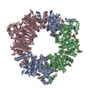 2btyC 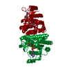 1gs5S S: Starting model for refinement C: citing same article ( |
|---|---|
| Similar structure data |


-Links
 PDBj
PDBj
- Assembly
| Deposited unit | 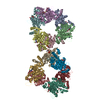
| ||||||||
|---|---|---|---|---|---|---|---|---|---|
| 1 | 
| ||||||||
| 2 | 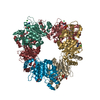
| ||||||||
| Unit cell |
|
-Components
| #1: Protein | / NAG KINASE / AGK / N-ACETYL-L-GLUTAMATE 5-PHOSPHOTRANSFERASE Mass: 31754.838 Da / Num. of mol.: 12 Source method: isolated from a genetically manipulated source Source: (gene. exp.) PSEUDOMONAS AERUGINOSA (bacteria) / Plasmid: PNAGK-PA25 / Production host: ESCHERICHIA COLI (E. coli) / Strain (production host): BL21(DE3) / References: UniProt: Q9HTN2, acetylglutamate kinase#2: Chemical | ChemComp-NLG / N-Acetylglutamic acid  Mass: 189.166 Da / Num. of mol.: 10 / Source method: obtained synthetically / Formula: C7H11NO5 Mass: 189.166 Da / Num. of mol.: 10 / Source method: obtained synthetically / Formula: C7H11NO5#3: Chemical | ChemComp-ADP / Adenosine diphosphate  Mass: 427.201 Da / Num. of mol.: 10 / Source method: obtained synthetically / Formula: C10H15N5O10P2 / Comment: ADP, energy-carrying molecule*YM Mass: 427.201 Da / Num. of mol.: 10 / Source method: obtained synthetically / Formula: C10H15N5O10P2 / Comment: ADP, energy-carrying molecule*YM#4: Chemical | ChemComp-MG /   Mass: 24.305 Da / Num. of mol.: 8 / Source method: obtained synthetically / Formula: Mg Mass: 24.305 Da / Num. of mol.: 8 / Source method: obtained synthetically / Formula: Mg#5: Chemical | ChemComp-CL / Chloride  Mass: 35.453 Da / Num. of mol.: 6 / Source method: obtained synthetically / Formula: Cl Mass: 35.453 Da / Num. of mol.: 6 / Source method: obtained synthetically / Formula: Cl |
|---|
-Experimental details
-Experiment
| Experiment | Method: X-RAY DIFFRACTION / Number of used crystals: 1 |
|---|
- Sample preparation
| Crystal | Density Matthews: 2.89 Å3/Da / Density % sol: 57.17 % |
|---|---|
| Crystal grow | Temperature: 277 K / Method: vapor diffusion, hanging drop / pH: 6.5 Details: CRYSTALS WERE OBTAINED AT 277 K IN ABOUT TWO WEEKS USING THE HANGING DROP TECHNIQUE AND UTILIZING A 10 MG/ML. PROTEIN SOLUTION CONTAINING 20 MM HEPES PH 7.5, 1 MM DITHIOERYTHRITOL, 10% ...Details: CRYSTALS WERE OBTAINED AT 277 K IN ABOUT TWO WEEKS USING THE HANGING DROP TECHNIQUE AND UTILIZING A 10 MG/ML. PROTEIN SOLUTION CONTAINING 20 MM HEPES PH 7.5, 1 MM DITHIOERYTHRITOL, 10% GLYCEROL, 30 MM MAGNESIUM CHLORIDE, 20 MM N-ACETYL-L-GLUTAMATE, 10 MM ADP AND 0.02% SODIUM AZIDE, AND A RESERVOIR SOLUTION WITH 0.1 M SODIUM CACODYLATE PH 6.5, 150-170 MM MAGNESIUM ACETATE AND POLYETHYLENE GLYCOL 8000 |
-Data collection
| Diffraction | Mean temperature: 100 K |
|---|---|
| Diffraction source | Source: SYNCHROTRON / Site: ESRF  / Beamline: ID14-4 / Wavelength: 0.973948 / Beamline: ID14-4 / Wavelength: 0.973948 |
| Detector | Type: ADSC CCD / Detector: CCD / Date: Feb 15, 2001 |
| Radiation | Protocol: SINGLE WAVELENGTH / Monochromatic (M) / Laue (L): M / Scattering type: x-ray |
| Radiation wavelength | Wavelength: 0.973948 Å / Relative weight: 1 |
| Reflection | Resolution: 2.95→69 Å / Num. obs: 86766 / % possible obs: 96.6 % / Redundancy: 1.9 % / Biso Wilson estimate: 86.056 Å2 / Rmerge(I) obs: 0.05 / Net I/σ(I): 5.4 |
| Reflection shell | Resolution: 2.95→3.1 Å / Rmerge(I) obs: 0.36 / Mean I/σ(I) obs: 2 / % possible all: 94.2 |
- Processing
| Software |
| ||||||||||||||||||||||||||||||||||||||||||||||||||||||||||||
|---|---|---|---|---|---|---|---|---|---|---|---|---|---|---|---|---|---|---|---|---|---|---|---|---|---|---|---|---|---|---|---|---|---|---|---|---|---|---|---|---|---|---|---|---|---|---|---|---|---|---|---|---|---|---|---|---|---|---|---|---|---|
| Refinement | Method to determine structure: MOLECULAR REPLACEMENT Starting model: PDB ENTRY 1GS5 Resolution: 2.95→18 Å / Data cutoff high absF: 10000 / Cross valid method: THROUGHOUT / σ(F): 0
| ||||||||||||||||||||||||||||||||||||||||||||||||||||||||||||
| Displacement parameters | Biso mean: 88.333 Å2
| ||||||||||||||||||||||||||||||||||||||||||||||||||||||||||||
| Refinement step | Cycle: LAST / Resolution: 2.95→18 Å
| ||||||||||||||||||||||||||||||||||||||||||||||||||||||||||||
| Refine LS restraints |
| ||||||||||||||||||||||||||||||||||||||||||||||||||||||||||||
| Xplor file |
|
