 Movie
Movie Controller

[English] 日本語
 Yorodumi
- PDB-2bqx: Inorganic Pyrophosphatase from the Pathogenic Bacterium Helicobac... -
+ Open data

- Basic information
| Entry | Database: PDB / ID: 2bqx | ||||||
|---|---|---|---|---|---|---|---|
| Title | Inorganic Pyrophosphatase from the Pathogenic Bacterium Helicobacter pylori-Kinetic and Structural Properties | ||||||
 Components Components | INORGANIC PYROPHOSPHATASE | ||||||
 Keywords Keywords | HYDROLASE / INORGANIC PYROPHOSPHATASE / HELICOBACTER PYLORI | ||||||
| Function / homology |  Function and homology informationinorganic diphosphatase / inorganic diphosphate phosphatase activity / phosphate-containing compound metabolic process / magnesium ion binding / cytosol Function and homology informationinorganic diphosphatase / inorganic diphosphate phosphatase activity / phosphate-containing compound metabolic process / magnesium ion binding / cytosolSimilarity search - Function | ||||||
| Biological species |   HELICOBACTER PYLORI (bacteria) HELICOBACTER PYLORI (bacteria) | ||||||
| Method | X-RAY DIFFRACTION / SYNCHROTRON / MOLECULAR REPLACEMENT / Resolution: 1.9 Å | ||||||
 Authors Authors | Chao, T.-C. / Sun, Y.-J. | ||||||
 Citation Citation | Journal: Proteins / Year: 2006 Title: Kinetic and Structural Properties of Inorganic Pyrophosphatase from the Pathogenic Bacterium Helicobacter Pylori. Authors: Chao, T.-C. / Huang, H. / Tsai, J.Y. / Huang, C.Y. / Sun, Y.-J. | ||||||
| History |
| ||||||
| Remark 700 | SHEET THE SHEET STRUCTURE OF THIS MOLECULE IS BIFURCATED. IN ORDER TO REPRESENT THIS FEATURE IN ... SHEET THE SHEET STRUCTURE OF THIS MOLECULE IS BIFURCATED. IN ORDER TO REPRESENT THIS FEATURE IN THE SHEET RECORDS BELOW, TWO SHEETS ARE DEFINED. |
- Structure visualization
| Structure viewer | Molecule: MolmilJmol/JSmol |
|---|
- Downloads & links
-Download
| PDBx/mmCIF format | 2bqx.cif.gz | 50.4 KB | Display | PDBx/mmCIF format |
|---|---|---|---|---|
| PDB format | pdb2bqx.ent.gz | 35.2 KB | Display | PDB format |
| PDBx/mmJSON format | 2bqx.json.gz | Tree view | PDBx/mmJSON format | |
| Others |  Other downloads Other downloads |
-Validation report
| Arichive directory | https://data.pdbj.org/pub/pdb/validation_reports/bq/2bqxftp://data.pdbj.org/pub/pdb/validation_reports/bq/2bqx | HTTPS FTP |
|---|
-Related structure data
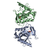
| Related structure data | 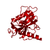 2bqyC  1qezS C: citing same article ( S: Starting model for refinement |
|---|---|
| Similar structure data |


-Links
 PDBj
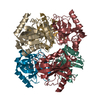
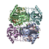
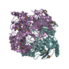
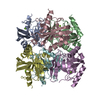
PDBj- Assembly
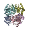
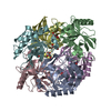
| Deposited unit | 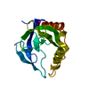
| |||||||||||||||
|---|---|---|---|---|---|---|---|---|---|---|---|---|---|---|---|---|
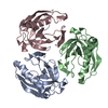
| 1 | x 6
| |||||||||||||||
| Unit cell |
| |||||||||||||||
| Components on special symmetry positions |
|
-Components
| #1: Protein | / PYROPHOSPHATE PHOSPHO-HYDROLASE / PPASE Mass: 19297.938 Da / Num. of mol.: 1 Source method: isolated from a genetically manipulated source Source: (gene. exp.) HELICOBACTER PYLORI (bacteria) / Strain: 26695 / Plasmid: PQE30 / Production host: ESCHERICHIA COLI (E. coli) / Strain (production host): SG13009 / References: UniProt: P56153, inorganic diphosphatase |
|---|---|
| #2: Water | ChemComp-HOH / Water Mass: 18.015 Da / Num. of mol.: 191 / Source method: isolated from a natural source / Formula: H2O Mass: 18.015 Da / Num. of mol.: 191 / Source method: isolated from a natural source / Formula: H2O |
-Experimental details
-Experiment
| Experiment | Method: X-RAY DIFFRACTION / Number of used crystals: 1 |
|---|
- Sample preparation
| Crystal | Density Matthews: 3.44 Å3/Da / Density % sol: 64 % |
|---|
-Data collection
| Diffraction | Mean temperature: 100 K |
|---|---|
| Diffraction source | Source: SYNCHROTRON / Site: SPring-8  / Beamline: BL12B2 / Wavelength: 0.9793 / Beamline: BL12B2 / Wavelength: 0.9793 |
| Detector | Type: ADSC CCD / Detector: CCD / Date: Dec 12, 2003 |
| Radiation | Protocol: SINGLE WAVELENGTH / Monochromatic (M) / Laue (L): M / Scattering type: x-ray |
| Radiation wavelength | Wavelength: 0.9793 Å / Relative weight: 1 |
| Reflection | Resolution: 1.9→19.93 Å / Num. obs: 192336 / % possible obs: 99.4 % / Observed criterion σ(I): 0 / Redundancy: 8.1 % / Biso Wilson estimate: 17.4 Å2 / Rmerge(I) obs: 0.1 |
| Reflection shell | Resolution: 1.9→2.02 Å / Redundancy: 5.2 % / Rmerge(I) obs: 0.36 / % possible all: 99.4 |
- Processing
| Software |
| ||||||||||||||||||||||||||||||||||||||||||||||||||||||||||||||||||||||||||||||||
|---|---|---|---|---|---|---|---|---|---|---|---|---|---|---|---|---|---|---|---|---|---|---|---|---|---|---|---|---|---|---|---|---|---|---|---|---|---|---|---|---|---|---|---|---|---|---|---|---|---|---|---|---|---|---|---|---|---|---|---|---|---|---|---|---|---|---|---|---|---|---|---|---|---|---|---|---|---|---|---|---|---|
| Refinement | Method to determine structure: MOLECULAR REPLACEMENT Starting model: PDB ENTRY 1QEZ Resolution: 1.9→19.93 Å / Rfactor Rfree error: 0.007 / Data cutoff high absF: 355817.05 / Isotropic thermal model: RESTRAINED / Cross valid method: THROUGHOUT / σ(F): 0
| ||||||||||||||||||||||||||||||||||||||||||||||||||||||||||||||||||||||||||||||||
| Solvent computation | Solvent model: FLAT MODEL / Bsol: 45.5299 Å2 / ksol: 0.384942 e/Å3 | ||||||||||||||||||||||||||||||||||||||||||||||||||||||||||||||||||||||||||||||||
| Displacement parameters | Biso mean: 30.1 Å2
| ||||||||||||||||||||||||||||||||||||||||||||||||||||||||||||||||||||||||||||||||
| Refine analyze |
| ||||||||||||||||||||||||||||||||||||||||||||||||||||||||||||||||||||||||||||||||
| Refinement step | Cycle: LAST / Resolution: 1.9→19.93 Å
| ||||||||||||||||||||||||||||||||||||||||||||||||||||||||||||||||||||||||||||||||
| Refine LS restraints |
| ||||||||||||||||||||||||||||||||||||||||||||||||||||||||||||||||||||||||||||||||
| LS refinement shell | Resolution: 1.9→2.02 Å / Rfactor Rfree error: 0.02 / Total num. of bins used: 6
| ||||||||||||||||||||||||||||||||||||||||||||||||||||||||||||||||||||||||||||||||
| Xplor file |
|
