 Movie
Movie Controller


+ Open data

- Basic information
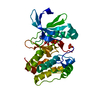
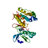
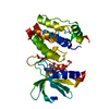
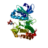
| Entry | Database: PDB / ID: 2bmc | ||||||
|---|---|---|---|---|---|---|---|
| Title | Aurora-2 T287D T288D complexed with PHA-680632 | ||||||
 Components Components | SERINE THREONINE-PROTEIN KINASE 6 | ||||||
 Keywords Keywords |  TRANSFERASE / CELL CYCLE / SERINE/THREONINE-PROTEIN KINASE / ATP-BINDING / PHOSPHORYLATION TRANSFERASE / CELL CYCLE / SERINE/THREONINE-PROTEIN KINASE / ATP-BINDING / PHOSPHORYLATION | ||||||
| Function / homology |  Function and homology information Function and homology informationInteraction between PHLDA1 and AURKA / regulation of centrosome cycle / axon hillock / spindle assembly involved in female meiosis I / cilium disassembly / spindle pole centrosome / positive regulation of oocyte maturation / histone H3S10 kinase activity / chromosome passenger complex / pronucleus ...Interaction between PHLDA1 and AURKA / regulation of centrosome cycle / axon hillock / spindle assembly involved in female meiosis I / cilium disassembly / spindle pole centrosome / positive regulation of oocyte maturation / histone H3S10 kinase activity / chromosome passenger complex / pronucleus / meiotic spindle / mitotic centrosome separation / germinal vesicle / protein localization to centrosome / anterior/posterior axis specification / centrosome localization / neuron projection extension / positive regulation of mitochondrial fission / spindle organization / mitotic spindle pole / SUMOylation of DNA replication proteins / spindle midzone / regulation of G2/M transition of mitotic cell cycle / centriole / protein serine/threonine/tyrosine kinase activity / positive regulation of mitotic cell cycle / AURKA Activation by TPX2 / mitotic spindle organization / TP53 Regulates Transcription of Genes Involved in G2 Cell Cycle Arrest / positive regulation of mitotic nuclear division / ciliary basal body / negative regulation of protein binding / regulation of signal transduction by p53 class mediator / regulation of cytokinesis / molecular function activator activity / liver regeneration / FBXL7 down-regulates AURKA during mitotic entry and in early mitosis / APC/C:Cdh1 mediated degradation of Cdc20 and other APC/C:Cdh1 targeted proteins in late mitosis/early G1 / regulation of protein stability / mitotic spindle / spindle / kinetochore / response to wounding / microtubule cytoskeleton / G2/M transition of mitotic cell cycle / Regulation of PLK1 Activity at G2/M Transition / positive regulation of proteasomal ubiquitin-dependent protein catabolic process / mitotic cell cycle / midbody / proteasome-mediated ubiquitin-dependent protein catabolic process / basolateral plasma membrane / peptidyl-serine phosphorylation / Regulation of TP53 Activity through Phosphorylation / microtubule / postsynaptic density / protein autophosphorylation / non-specific serine/threonine protein kinase / protein kinase activity / protein heterodimerization activity / cell division / negative regulation of gene expression / protein phosphorylation / protein serine kinase activity / centrosome / protein serine/threonine kinase activity / glutamatergic synapse / apoptotic process / ubiquitin protein ligase binding / negative regulation of apoptotic process / protein kinase binding / perinuclear region of cytoplasm / nucleoplasm / ATP binding / nucleus / cytosolSimilarity search - Function | ||||||
| Biological species |  HOMO SAPIENS (human) HOMO SAPIENS (human) | ||||||
| Method | X-RAY DIFFRACTION / SYNCHROTRON / MOLECULAR REPLACEMENT / Resolution: 2.6 Å | ||||||
 Authors Authors | Cameron, A.D. / Izzo, G. / Sagliano, A. / Rusconi, L. / Storici, P. / Fancelli, D. / Berta, D. / Bindi, S. / Catana, C. / Forte, B. ...Cameron, A.D. / Izzo, G. / Sagliano, A. / Rusconi, L. / Storici, P. / Fancelli, D. / Berta, D. / Bindi, S. / Catana, C. / Forte, B. / Giordano, P. / Mantegani, S. / Meroni, M. / Moll, J. / Pittala, V. / Severino, D. / Tonani, R. / Varasi, M. / Vulpetti, A. / Vianello, P. | ||||||
 Citation Citation | Journal: J.Med.Chem. / Year: 2005 Title: Potent and Selective Aurora Inhibitors Identified by the Expansion of a Novel Scaffold for Protein Kinase Inhibition. Authors: Fancelli, D. / Berta, D. / Bindi, S. / Cameron, A. / Cappella, P. / Carpinelli, P. / Catana, C. / Forte, B. / Giordano, P. / Giorgini, M.L. / Mantegani, S. / Marsiglio, A. / Meroni, M. / ...Authors: Fancelli, D. / Berta, D. / Bindi, S. / Cameron, A. / Cappella, P. / Carpinelli, P. / Catana, C. / Forte, B. / Giordano, P. / Giorgini, M.L. / Mantegani, S. / Marsiglio, A. / Meroni, M. / Moll, J. / Pittala, V. / Roletto, F. / Severino, D. / Soncini, C. / Storici, P. / Tonani, R. / Varasi, M. / Vulpetti, A. / Vianello, P. | ||||||
| History |
|
- Structure visualization
| Structure viewer | Molecule: MolmilJmol/JSmol |
|---|
- Downloads & links
-Download
| PDBx/mmCIF format | 2bmc.cif.gz | 322.2 KB | Display | PDBx/mmCIF format |
|---|---|---|---|---|
| PDB format | pdb2bmc.ent.gz | 272.4 KB | Display | PDB format |
| PDBx/mmJSON format | 2bmc.json.gz | Tree view | PDBx/mmJSON format | |
| Others |  Other downloads Other downloads |
-Validation report
| Arichive directory | https://data.pdbj.org/pub/pdb/validation_reports/bm/2bmcftp://data.pdbj.org/pub/pdb/validation_reports/bm/2bmc | HTTPS FTP |
|---|
-Related structure data
| Related structure data | |
|---|---|
| Similar structure data |


-Links
 PDBj
PDBj


- Assembly
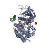
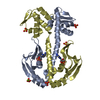
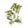
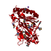
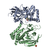
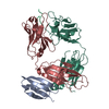
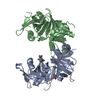
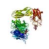
| Deposited unit | 
| ||||||||||||||||||||||||
|---|---|---|---|---|---|---|---|---|---|---|---|---|---|---|---|---|---|---|---|---|---|---|---|---|---|
| 1 | 
| ||||||||||||||||||||||||
| 2 | 
| ||||||||||||||||||||||||
| 3 | 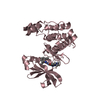
| ||||||||||||||||||||||||
| 4 | 
| ||||||||||||||||||||||||
| 5 | 
| ||||||||||||||||||||||||
| 6 | 
| ||||||||||||||||||||||||
| Unit cell |
| ||||||||||||||||||||||||
| Noncrystallographic symmetry (NCS) | NCS oper:
|
-Components
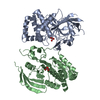
| #1: Protein | Mass: 35305.164 Da / Num. of mol.: 6 / Fragment: CATALYTIC DOMAIN, RESIDUES 100-403 / Mutation: YES Source method: isolated from a genetically manipulated source Source: (gene. exp.) HOMO SAPIENS (human) / Cell line (production host): High Five / Production host:  TRICHOPLUSIA NI (cabbage looper) / References: UniProt: O14965, EC: 2.7.1.37 TRICHOPLUSIA NI (cabbage looper) / References: UniProt: O14965, EC: 2.7.1.37#2: Chemical | ChemComp-MPY / (   Mass: 497.591 Da / Num. of mol.: 6 / Source method: obtained synthetically / Formula: C28H31N7O2 Mass: 497.591 Da / Num. of mol.: 6 / Source method: obtained synthetically / Formula: C28H31N7O2#3: Water | ChemComp-HOH / | Water Mass: 18.015 Da / Num. of mol.: 200 / Source method: isolated from a natural source / Formula: H2O Mass: 18.015 Da / Num. of mol.: 200 / Source method: isolated from a natural source / Formula: H2OCompound details | FUNCTION: POSSIBLE ROLE IN CELL CYCLE REGULATION DURING ANAPHASE AND/OR TELOPHASE ENGINEERED ...FUNCTION: POSSIBLE ROLE IN CELL CYCLE REGULATION | Sequence details | ADDITIONAL 2 AMINO ACIDS AT N-TERMINUS DUE TO CLONING ARTIFACT. THE MUTATION IN THIS STRUCTURE THR ...ADDITIONAL | |
|---|
-Experimental details
-Experiment
| Experiment | Method: X-RAY DIFFRACTION / Number of used crystals: 1 |
|---|
- Sample preparation
| Crystal | Density Matthews: 3.62 Å3/Da / Density % sol: 65.78 % |
|---|---|
| Crystal grow | pH: 4.6 Details: 2.5M NACL, 0.1M NAAC, 0.2M LISO4 WITH NON-DETERGENT SULFOBETAINE 195 AS ADDITIVE IN THE DROP, pH 4.60 |
-Data collection
| Diffraction | Mean temperature: 100 K |
|---|---|
| Diffraction source | Source: SYNCHROTRON / Site: ESRF  / Beamline: ID14-1 / Wavelength: 0.93 / Beamline: ID14-1 / Wavelength: 0.93 |
| Detector | Type: ADSC CCD / Detector: CCD / Date: Jan 25, 2002 |
| Radiation | Monochromator: DIAMOND (111), GE(220) / Protocol: SINGLE WAVELENGTH / Monochromatic (M) / Laue (L): M / Scattering type: x-ray |
| Radiation wavelength | Wavelength: 0.93 Å / Relative weight: 1 |
| Reflection | Resolution: 2.6→40 Å / Num. obs: 77069 / % possible obs: 97.4 % / Observed criterion σ(I): 2 / Redundancy: 2.4 % / Biso Wilson estimate: 47.1 Å2 / Rmerge(I) obs: 0.08 / Net I/σ(I): 6.9 |
| Reflection shell | Resolution: 2.6→2.74 Å / Redundancy: 2.4 % / Rmerge(I) obs: 0.33 / Mean I/σ(I) obs: 2.1 / % possible all: 97.4 |
- Processing
| Software |
| ||||||||||||||||||||||||||||||||||||||||||||||||||||||||||||||||||||||||||||||||
|---|---|---|---|---|---|---|---|---|---|---|---|---|---|---|---|---|---|---|---|---|---|---|---|---|---|---|---|---|---|---|---|---|---|---|---|---|---|---|---|---|---|---|---|---|---|---|---|---|---|---|---|---|---|---|---|---|---|---|---|---|---|---|---|---|---|---|---|---|---|---|---|---|---|---|---|---|---|---|---|---|---|
| Refinement | Method to determine structure: MOLECULAR REPLACEMENT Starting model: UNPUBLISHED STRUCTURE Resolution: 2.6→29.95 Å / Rfactor Rfree error: 0.004 / Isotropic thermal model: RESTRAINED / Cross valid method: THROUGHOUT / σ(F): 0 Details: BULK SOLVENT MODEL USED. THE 6 MOLECULES OF THE ASYMMETRIC UNIT ARE ARRANGED IN 3 PAIRS OF DIMERS. WITHIN THESE DIMERS DOMAIN SWAPPING IS CLEARLY EVIDENT INVOLVING RESIDUES 292 TO 306 ...Details: BULK SOLVENT MODEL USED. THE 6 MOLECULES OF THE ASYMMETRIC UNIT ARE ARRANGED IN 3 PAIRS OF DIMERS. WITHIN THESE DIMERS DOMAIN SWAPPING IS CLEARLY EVIDENT INVOLVING RESIDUES 292 TO 306 ALTHOUGH FROM RESIDUES 304 TO 306 THE DENSITY IS RATHER POOR. RESIDUES 280-291 IN THE ACTIVATION LOOP BEFORE THIS REGION CANNOT BE OBSERVED
| ||||||||||||||||||||||||||||||||||||||||||||||||||||||||||||||||||||||||||||||||
| Solvent computation | Solvent model: FLAT MODEL / Bsol: 36.5298 Å2 / ksol: 0.369773 e/Å3 | ||||||||||||||||||||||||||||||||||||||||||||||||||||||||||||||||||||||||||||||||
| Displacement parameters | Biso mean: 48.7 Å2
| ||||||||||||||||||||||||||||||||||||||||||||||||||||||||||||||||||||||||||||||||
| Refine analyze |
| ||||||||||||||||||||||||||||||||||||||||||||||||||||||||||||||||||||||||||||||||
| Refinement step | Cycle: LAST / Resolution: 2.6→29.95 Å
| ||||||||||||||||||||||||||||||||||||||||||||||||||||||||||||||||||||||||||||||||
| Refine LS restraints |
| ||||||||||||||||||||||||||||||||||||||||||||||||||||||||||||||||||||||||||||||||
| Refine LS restraints NCS | NCS model details: RESTRAINTS / Rms dev Biso : 1.5 Å2 / Rms dev position: 0.5 Å / Weight Biso : 2 / Weight position: 10 | ||||||||||||||||||||||||||||||||||||||||||||||||||||||||||||||||||||||||||||||||
| LS refinement shell | Resolution: 2.6→2.76 Å / Rfactor Rfree error: 0.015 / Total num. of bins used: 6
| ||||||||||||||||||||||||||||||||||||||||||||||||||||||||||||||||||||||||||||||||
| Xplor file | Serial no: 1 / Param file: PROTEIN_REP.PARAM / Topol file: 632.TOP |
