 Movie
Movie Controller

[English] 日本語
 Yorodumi
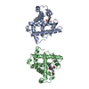
- PDB-2bgz: ATOMIC MODEL OF THE BACTERIAL FLAGELLAR BASED ON DOCKING AN X-RAY... -
+ Open data

- Basic information
| Entry | Database: PDB / ID: 2bgz | ||||||
|---|---|---|---|---|---|---|---|
| Title | ATOMIC MODEL OF THE BACTERIAL FLAGELLAR BASED ON DOCKING AN X-RAY DERIVED HOOK STRUCTURE INTO AN EM MAP. | ||||||
 Components Components | FLAGELLAR HOOK PROTEIN FLGE | ||||||
 Keywords Keywords |  MOTOR PROTEIN / BACTERIAL MOTIILITY / BACTERIAL FLAGELLAR HOOK / FLAGELLUM MOTOR PROTEIN / BACTERIAL MOTIILITY / BACTERIAL FLAGELLAR HOOK / FLAGELLUM | ||||||
| Function / homology |  Function and homology information Function and homology informationbacterial-type flagellum basal body / bacterial-type flagellum-dependent swarming motility Similarity search - Function | ||||||
| Biological species |  SALMONELLA TYPHIMURIUM (bacteria) SALMONELLA TYPHIMURIUM (bacteria) | ||||||
| Method | ELECTRON MICROSCOPY / electron crystallography / cryo EM / Resolution: 9 Å | ||||||
 Authors Authors | Shaikh, T.R. / Thomas, D.R. / Chen, J.Z. / Samatey, F.A. / Matsunami, H. / Imada, K. / Namba, K. / Derosier, D.J. | ||||||
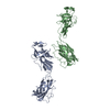
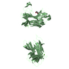
 Citation Citation | Journal: Proc Natl Acad Sci U S A / Year: 2005 Title: A partial atomic structure for the flagellar hook of Salmonella typhimurium. Authors: Tanvir R Shaikh / Dennis R Thomas / James Z Chen / Fadel A Samatey / Hideyuki Matsunami / Katsumi Imada / Keiichi Namba / David J Derosier /  Abstract: The axial proteins of the bacterial flagellum function as a drive shaft, universal joint, and propeller driven by the flagellar rotary motor; they also form the putative protein export channel. The N- ...The axial proteins of the bacterial flagellum function as a drive shaft, universal joint, and propeller driven by the flagellar rotary motor; they also form the putative protein export channel. The N- and C-terminal sequences of the eight axial proteins were predicted to form interlocking alpha-domains generating an axial tube. We report on an approximately 1-nm resolution map of the hook from Salmonella typhimurium, which reveals such a tube made from interdigitated, 1-nm rod-like densities similar to those seen in maps of the filament. Atomic models for the two outer domains of the hook subunit were docked into the corresponding outermost features of the map. The N and C termini of the hook subunit fragment are positioned next to each other and face toward the axis of the hook. The placement of these termini would permit the residues missing in the fragment to form the rod-like features that form the core domain of the hook. We also fit the hook atomic model to an approximately 2-nm resolution map of the hook from Caulobacter crescentus. The hook protein sequence from C. crescentus is largely homologous to that of S. typhimurium except for a large insertion (20 kDa). According to difference maps and our fitting, this insertion is found on the outer surface of the hook, consistent with our modeling of the hook. #1: Journal: Nature / Year: 2004Title: Structure of the bacterial flagellar hook and implication for the molecular universal joint mechanism. Authors: Fadel A Samatey / Hideyuki Matsunami / Katsumi Imada / Shigehiro Nagashima / Tanvir R Shaikh / Dennis R Thomas / James Z Chen / David J Derosier / Akio Kitao / Keiichi Namba / Abstract: The bacterial flagellum is a motile organelle, and the flagellar hook is a short, highly curved tubular structure that connects the flagellar motor to the long filament acting as a helical propeller. ...The bacterial flagellum is a motile organelle, and the flagellar hook is a short, highly curved tubular structure that connects the flagellar motor to the long filament acting as a helical propeller. The hook is made of about 120 copies of a single protein, FlgE, and its function as a nano-sized universal joint is essential for dynamic and efficient bacterial motility and taxis. It transmits the motor torque to the helical propeller over a wide range of its orientation for swimming and tumbling. Here we report a partial atomic model of the hook obtained by X-ray crystallography of FlgE31, a major proteolytic fragment of FlgE lacking unfolded terminal regions, and by electron cryomicroscopy and three-dimensional helical image reconstruction of the hook. The model reveals the intricate molecular interactions and a plausible switching mechanism for the hook to be flexible in bending but rigid against twisting for its universal joint function. | ||||||
| History |
| ||||||
| Remark 700 | SHEET THE SHEET STRUCTURE OF THIS MOLECULE IS BIFURCATED. IN ORDER TO REPRESENT THIS FEATURE IN ... SHEET THE SHEET STRUCTURE OF THIS MOLECULE IS BIFURCATED. IN ORDER TO REPRESENT THIS FEATURE IN THE SHEET RECORDS BELOW, TWO SHEETS ARE DEFINED. |
- Structure visualization
| Movie |
Movie viewer |
|---|---|
| Structure viewer | Molecule: MolmilJmol/JSmol |
- Downloads & links
-Download
| PDBx/mmCIF format | 2bgz.cif.gz | 61.9 KB | Display | PDBx/mmCIF format |
|---|---|---|---|---|
| PDB format | pdb2bgz.ent.gz | 47.7 KB | Display | PDB format |
| PDBx/mmJSON format | 2bgz.json.gz | Tree view | PDBx/mmJSON format | |
| Others |  Other downloads Other downloads |
-Validation report
| Arichive directory | https://data.pdbj.org/pub/pdb/validation_reports/bg/2bgzftp://data.pdbj.org/pub/pdb/validation_reports/bg/2bgz | HTTPS FTP |
|---|
-Related structure data
| Related structure data | 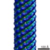 1132MUC 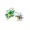 2bgyC C: citing same article ( M: map data used to model this data U: unfit; in different coordinate system*YM |
|---|---|
| Similar structure data |


-Links
 PDBj
PDBj
- Assembly
| Deposited unit | 
|
|---|---|
| 1 |
|
-Components
| #1: Protein | Mass: 31335.084 Da / Num. of mol.: 1 / Fragment: FLGE31, RESIDUES 71-369 Source method: isolated from a genetically manipulated source Details: FIT OF CRYSTAL STRUCTURE PDB ID 1WLG TO AN EM AMAP OF S. TYPHIMURIUM POLYHOOK Source: (gene. exp.) SALMONELLA TYPHIMURIUM (bacteria) / Production host: ESCHERICHIA COLI (E. coli) / References: UniProt: P16322, UniProt: P0A1J1*PLUS |
|---|
-Experimental details
-Experiment
| Experiment | Method: ELECTRON MICROSCOPY |
|---|---|
| EM experiment | Aggregation state: 2D ARRAY / 3D reconstruction method: electron crystallography |
- Sample preparation
| Component | Name: POLYHOOK / Type: COMPLEX |
|---|---|
| Buffer solution | Name: 10 MM TRIS/HCL,5MM EDTA,0. 1%V/V TRITON X-100 / pH: 8 / Details: 10 MM TRIS/HCL,5MM EDTA,0. 1%V/V TRITON X-100 |
| Specimen | Embedding applied: NO / Shadowing applied: NO / Staining applied: NO / Vitrification applied: YES |
| Specimen support | Details: HOLEY CARBON |
| Vitrification | Instrument: HOMEMADE PLUNGER / Cryogen name: ETHANE Details: SAMPLES PREPARED AT 4 DEG C PLUNGED INTO LIQUID ETHANE |
- Electron microscopy imaging
| Microscopy | Model: FEI/PHILIPS CM200FEG Details: DATA FROM 262 INDIVIDUAL SEGMENTS OF HOOK IN FINAL MAP. |
|---|---|
| Electron gun | Electron source: FIELD EMISSION GUN / Accelerating voltage: 200 kV / Illumination mode: FLOOD BEAM |
| Electron lens | Mode: BRIGHT FIELDBright-field microscopy / Nominal magnification: 66000 X / Nominal defocus max: 2700 nm / Nominal defocus min: 1300 nm / Cs: 2 mm |
| Image recording | Electron dose: 10 e/Å2 / Film or detector model: KODAK SO-163 FILM |
| Radiation wavelength | Relative weight: 1 |
- Processing
| EM software |
| ||||||||||||
|---|---|---|---|---|---|---|---|---|---|---|---|---|---|
| 3D reconstruction | Resolution: 9 Å / Resolution method: DIFFRACTION PATTERN/LAYERLINES Details: HELICAL SYMMETRY OPERATORS CAN BE CALCULATED USING RISE PER SUBUNIT 4.226475A ROTATION PER SUBUNIT 23.48758 DEG. DATA USED IN REFINEMENT. RESOLUTION RANGE HIGH 12A, RESOLUTION RANGE LOW ...Details: HELICAL SYMMETRY OPERATORS CAN BE CALCULATED USING RISE PER SUBUNIT 4.226475A ROTATION PER SUBUNIT 23.48758 DEG. DATA USED IN REFINEMENT. RESOLUTION RANGE HIGH 12A, RESOLUTION RANGE LOW 200A, DATA CUTOFF (SIGMA(F)) 1, COMPLETENESS FOR RANGE 100(%) NUMBER OF REFLECTIONS 84. PROTEIN ATOMS 2156 Symmetry type: HELICAL | ||||||||||||
| Atomic model building | Protocol: OTHER / Details: METHOD--RECIPROCAL SPACE FITTING IN URO | ||||||||||||
| Refinement | Highest resolution: 12 Å | ||||||||||||
| Refinement step | Cycle: LAST / Highest resolution: 12 Å
|
