 Movie
Movie Controller

[English] 日本語
 Yorodumi
- PDB-2bgi: X-Ray Structure of the Ferredoxin-NADP(H) Reductase from Rhodobac... -
+ Open data

- Basic information
| Entry | Database: PDB / ID: 2bgi | ||||||
|---|---|---|---|---|---|---|---|
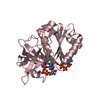
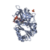
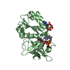
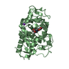
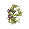
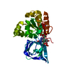
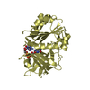
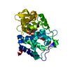
| Title | X-Ray Structure of the Ferredoxin-NADP(H) Reductase from Rhodobacter capsulatus complexed with three molecules of the detergent n-heptyl- beta-D-thioglucoside at 1.7 Angstroms | ||||||
 Components Components | FERREDOXIN-NADP(H) REDUCTASE | ||||||
 Keywords Keywords |  OXIDOREDUCTASE / FERREDOXIN(FLAVODOXIN)-NADP(H) REDUCTASE / FLAVOPROTEINS / ELECTRON TRANSFER / RHODOBACTER CAPSULATUS OXIDOREDUCTASE / FERREDOXIN(FLAVODOXIN)-NADP(H) REDUCTASE / FLAVOPROTEINS / ELECTRON TRANSFER / RHODOBACTER CAPSULATUS | ||||||
| Function / homology |  Function and homology information Function and homology informationflavodoxin-NADP+ reductase / ferredoxin-NADP+ reductase / ferredoxin-NADP+ reductase activity / nucleotide binding / cytoplasmSimilarity search - Function | ||||||
| Biological species |  RHODOBACTER CAPSULATUS (bacteria) RHODOBACTER CAPSULATUS (bacteria) | ||||||
| Method | X-RAY DIFFRACTION / SYNCHROTRON / MOLECULAR REPLACEMENT / Resolution: 1.68 Å | ||||||
 Authors Authors | Perez-Dorado, J.I. / Hermoso, J.A. / Nogues, I. / Frago, S. / Bittel, C. / Mayhew, S.G. / Gomez-Moreno, C. / Medina, M. / Cortez, N. / Carrillo, N. | ||||||
 Citation Citation | Journal: Biochemistry / Year: 2005 Title: The Ferredoxin-Nadp(H) Reductase from Rhodobacter Capsulatus: Molecular Structure and Catalytic Mechanism Authors: Nogues, I. / Perez-Dorado, J.I. / Frago, S. / Bittel, C. / Mayhew, S.G. / Gomez-Moreno, C. / Hermoso, J.A. / Medina, M. / Cortez, N. / Carrillo, N. #1: Journal: Acta Crystallogr.,Sect.D / Year: 2004 Title: Crystallization and Preliminary X-Ray Diffraction Analysis of Ferredoxin-Nadp(H) Reductase from Rhodobacter Capsulatus Authors: Perez-Dorado, J.I. / Bittel, C. / Cortez, N. / Hermoso, J.A. #2: Journal: FEBS Lett. / Year: 2003 Title: The Oxidant-Responsive Diaphorase of Rhodobacter Capsulatus is a Ferredoxin (Flavodoxin)-Nadp(H) Reductase Authors: Bittel, C. / Tabares, L.C. / Armesto, M. / Carrillo, N. / Cortez, N. | ||||||
| History |
|
- Structure visualization
| Structure viewer | Molecule: MolmilJmol/JSmol |
|---|
- Downloads & links
-Download
| PDBx/mmCIF format | 2bgi.cif.gz | 75.6 KB | Display | PDBx/mmCIF format |
|---|---|---|---|---|
| PDB format | pdb2bgi.ent.gz | 54.9 KB | Display | PDB format |
| PDBx/mmJSON format | 2bgi.json.gz | Tree view | PDBx/mmJSON format | |
| Others |  Other downloads Other downloads |
-Validation report
| Arichive directory | https://data.pdbj.org/pub/pdb/validation_reports/bg/2bgiftp://data.pdbj.org/pub/pdb/validation_reports/bg/2bgi | HTTPS FTP |
|---|
-Related structure data
| Related structure data |  2bgjC 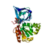 1a8pS C: citing same article ( S: Starting model for refinement |
|---|---|
| Similar structure data |


-Links
 PDBj
PDBj


- Assembly
| Deposited unit | 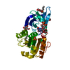
| ||||||||
|---|---|---|---|---|---|---|---|---|---|
| 1 |
| ||||||||
| Unit cell |
|
-Components
-Protein / Sugars , 2 types, 4 molecules A
| #1: Protein | Mass: 30436.889 Da / Num. of mol.: 1 / Mutation: YES Source method: isolated from a genetically manipulated source Source: (gene. exp.) RHODOBACTER CAPSULATUS (bacteria) / Strain: 37B4 / Variant: DSM938 / Production host: ESCHERICHIA COLI (E. coli) / Strain (production host): BL21 / References: UniProt: Q9L6V3, ferredoxin-NADP+ reductase |
|---|---|
| #3: Sugar |  Type: D-saccharide / Mass: 294.408 Da / Num. of mol.: 3 / Source method: obtained synthetically / Formula: C13H26O5S / Comment: detergent*YM Type: D-saccharide / Mass: 294.408 Da / Num. of mol.: 3 / Source method: obtained synthetically / Formula: C13H26O5S / Comment: detergent*YM |
-Non-polymers , 4 types, 268 molecules 


| #2: Chemical | ChemComp-FAD / Flavin adenine dinucleotide Mass: 785.550 Da / Num. of mol.: 1 / Source method: obtained synthetically / Formula: C27H33N9O15P2 / Comment: FAD*YM Mass: 785.550 Da / Num. of mol.: 1 / Source method: obtained synthetically / Formula: C27H33N9O15P2 / Comment: FAD*YM | ||
|---|---|---|---|
| #4: Chemical | ChemComp-SO4 / Sulfate Mass: 96.063 Da / Num. of mol.: 1 / Source method: obtained synthetically / Formula: SO4 Mass: 96.063 Da / Num. of mol.: 1 / Source method: obtained synthetically / Formula: SO4 | ||
| #5: Chemical | Carbon dioxide Mass: 44.010 Da / Num. of mol.: 2 / Source method: obtained synthetically / Formula: CO2 Mass: 44.010 Da / Num. of mol.: 2 / Source method: obtained synthetically / Formula: CO2#6: Water | ChemComp-HOH / | WaterMass: 18.015 Da / Num. of mol.: 264 / Source method: isolated from a natural source / Formula: H2O |
-Experimental details
-Experiment
| Experiment | Method: X-RAY DIFFRACTION / Number of used crystals: 1 |
|---|
- Sample preparation
| Crystal | Density Matthews: 3.56 Å3/Da / Density % sol: 64 % |
|---|---|
| Crystal grow | pH: 6.5 / Details: pH 6.50 |
-Data collection
| Diffraction | Mean temperature: 100 K |
|---|---|
| Diffraction source | Source: SYNCHROTRON / Site: ESRF  / Beamline: ID14-2 / Wavelength: 0.934 / Beamline: ID14-2 / Wavelength: 0.934 |
| Detector | Type: ADSC CCD / Detector: CCD / Date: May 17, 2004 |
| Radiation | Protocol: SINGLE WAVELENGTH / Monochromatic (M) / Laue (L): M / Scattering type: x-ray |
| Radiation wavelength | Wavelength: 0.934 Å / Relative weight: 1 |
| Reflection | Resolution: 1.7→45.6 Å / Num. obs: 61984 / % possible obs: 97.2 % / Observed criterion σ(I): 4 / Redundancy: 7.3 % / Biso Wilson estimate: 19.7 Å2 / Rmerge(I) obs: 0.12 / Net I/σ(I): 11.2 |
| Reflection shell | Resolution: 1.7→1.8 Å / Redundancy: 7.5 % / Rmerge(I) obs: 0.65 / Mean I/σ(I) obs: 2.6 / % possible all: 90 |
- Processing
| Software |
| ||||||||||||||||||||||||||||||||||||||||||||||||||||||||||||
|---|---|---|---|---|---|---|---|---|---|---|---|---|---|---|---|---|---|---|---|---|---|---|---|---|---|---|---|---|---|---|---|---|---|---|---|---|---|---|---|---|---|---|---|---|---|---|---|---|---|---|---|---|---|---|---|---|---|---|---|---|---|
| Refinement | Method to determine structure: MOLECULAR REPLACEMENT Starting model: PDB ENTRY 1A8P Resolution: 1.68→45.64 Å / Rfactor Rfree error: 0.004 / Data cutoff high absF: 1767576.25 / Isotropic thermal model: RESTRAINED / Cross valid method: THROUGHOUT / σ(F): 0 Details: RESIDUES 1 - 15 HAVE NOT BEEN MODELED BECAUSE THEY ARE NOT DEFINED IN THE ELECTRON DENSITY MAP
| ||||||||||||||||||||||||||||||||||||||||||||||||||||||||||||
| Solvent computation | Solvent model: FLAT MODEL / Bsol: 51.8594 Å2 / ksol: 0.389103 e/Å3 | ||||||||||||||||||||||||||||||||||||||||||||||||||||||||||||
| Displacement parameters | Biso mean: 25.3 Å2
| ||||||||||||||||||||||||||||||||||||||||||||||||||||||||||||
| Refine analyze |
| ||||||||||||||||||||||||||||||||||||||||||||||||||||||||||||
| Refinement step | Cycle: LAST / Resolution: 1.68→45.64 Å
| ||||||||||||||||||||||||||||||||||||||||||||||||||||||||||||
| Refine LS restraints |
| ||||||||||||||||||||||||||||||||||||||||||||||||||||||||||||
| LS refinement shell | Resolution: 1.68→1.79 Å / Rfactor Rfree error: 0.023 / Total num. of bins used: 6
|
