 Movie
Movie Controller

[English] 日本語
 Yorodumi
- PDB-2bdw: Crystal Structure of the Auto-Inhibited Kinase Domain of Calcium/... -
+ Open data

- Basic information
| Entry | Database: PDB / ID: 2bdw | ||||||
|---|---|---|---|---|---|---|---|
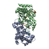
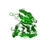
| Title | Crystal Structure of the Auto-Inhibited Kinase Domain of Calcium/Calmodulin Activated Kinase II | ||||||
 Components Components | Hypothetical protein K11E8.1d Hypothesis Hypothesis | ||||||
 Keywords Keywords | TRANSFERASE / kinase / calmodulin activated | ||||||
| Function / homology |  Function and homology information Function and homology informationmedium-term memory / Unblocking of NMDA receptors, glutamate binding and activation / Ion transport by P-type ATPases / Ion homeostasis / Ca2+ pathway / HSF1-dependent transactivation / serotonin biosynthetic process / Ca2+/calmodulin-dependent protein kinase / calmodulin-dependent protein kinase activity / axon cytoplasm ...medium-term memory / Unblocking of NMDA receptors, glutamate binding and activation / Ion transport by P-type ATPases / Ion homeostasis / Ca2+ pathway / HSF1-dependent transactivation / serotonin biosynthetic process / Ca2+/calmodulin-dependent protein kinase / calmodulin-dependent protein kinase activity / axon cytoplasm / peptidyl-threonine phosphorylation / MAPK cascade / perikaryon / peptidyl-serine phosphorylation / transmembrane transporter binding / calmodulin binding / neuron projection / phosphorylation / protein phosphorylation / protein serine kinase activity / positive regulation of gene expression / ATP binding / identical protein binding / metal ion binding / cytoplasmSimilarity search - Function | ||||||
| Biological species |  Caenorhabditis elegans (invertebrata) Caenorhabditis elegans (invertebrata) | ||||||
| Method | X-RAY DIFFRACTION / SYNCHROTRON / MOLECULAR REPLACEMENT / Resolution: 1.8 Å | ||||||
 Authors Authors | Rosenberg, O.S. / Kuriyan, J. | ||||||
 Citation Citation | Journal: Cell(Cambridge,Mass.) / Year: 2005 Title: Structure of the Autoinhibited Kinase Domain of CaMKII and SAXS Analysis of the Holoenzyme Authors: Rosenberg, O.S. / Deindl, S. / Sung, R.-J. / Nairn, A.C. / Kuriyan, J. | ||||||
| History |
|
- Structure visualization
| Structure viewer | Molecule: MolmilJmol/JSmol |
|---|
- Downloads & links
-Download
| PDBx/mmCIF format | 2bdw.cif.gz | 143.8 KB | Display | PDBx/mmCIF format |
|---|---|---|---|---|
| PDB format | pdb2bdw.ent.gz | 111.5 KB | Display | PDB format |
| PDBx/mmJSON format | 2bdw.json.gz | Tree view | PDBx/mmJSON format | |
| Others |  Other downloads Other downloads |
-Validation report
| Arichive directory | https://data.pdbj.org/pub/pdb/validation_reports/bd/2bdwftp://data.pdbj.org/pub/pdb/validation_reports/bd/2bdw | HTTPS FTP |
|---|
-Related structure data
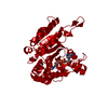
| Related structure data | 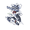 2phkS S: Starting model for refinement |
|---|---|
| Similar structure data |


-Links
 PDBj
PDBj
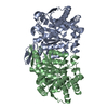
- Assembly
| Deposited unit | 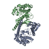
| ||||||||
|---|---|---|---|---|---|---|---|---|---|
| 1 |
| ||||||||
| Unit cell |
|
-Components
| #1: Protein | Hypothesis Mass: 40629.309 Da / Num. of mol.: 2 / Fragment: auto-inhibited kinase domain fragment / Mutation: D135N Source method: isolated from a genetically manipulated source Source: (gene. exp.) Caenorhabditis elegans (invertebrata)Gene: Ca2+/calmodulin-dependent protein kinase; unc-43 (K11E8.1d) Plasmid: pET28 / Species (production host): Escherichia coli / Production host:  Escherichia coli BL21(DE3) (bacteria) / Strain (production host): BL-21(DE3) / References: UniProt: Q9NG91, UniProt: O62305*PLUS Escherichia coli BL21(DE3) (bacteria) / Strain (production host): BL-21(DE3) / References: UniProt: Q9NG91, UniProt: O62305*PLUS#2: Water | ChemComp-HOH / | Water Mass: 18.015 Da / Num. of mol.: 431 / Source method: isolated from a natural source / Formula: H2O Mass: 18.015 Da / Num. of mol.: 431 / Source method: isolated from a natural source / Formula: H2O |
|---|
-Experimental details
-Experiment
| Experiment | Method: X-RAY DIFFRACTION / Number of used crystals: 1 |
|---|
- Sample preparation
| Crystal | Density Matthews: 2.6 Å3/Da / Density % sol: 52.62 % |
|---|---|
| Crystal grow | Temperature: 293 K / Method: vapor diffusion / pH: 7 Details: 1.6 M sodium malonate, 1.5 % 1,2,3-heptanetriol, pH 7.0, VAPOR DIFFUSION, temperature 293K |
-Data collection
| Diffraction | Mean temperature: 293 K |
|---|---|
| Diffraction source | Source: SYNCHROTRON / Site: ALS  / Beamline: 8.2.2 / Wavelength: 0.9537 Å / Beamline: 8.2.2 / Wavelength: 0.9537 Å |
| Detector | Type: ADSC QUANTUM 4 / Detector: CCD / Date: Dec 18, 2004 |
| Radiation | Monochromator: Double crystal, Si(111) / Protocol: SINGLE WAVELENGTH / Monochromatic (M) / Laue (L): M / Scattering type: x-ray |
| Radiation wavelength | Wavelength: 0.9537 Å / Relative weight: 1 |
| Reflection | Resolution: 1.8→45.8 Å / Num. all: 76218 / Num. obs: 73357 / % possible obs: 99.4 % / Observed criterion σ(F): 2 / Observed criterion σ(I): 1 / Biso Wilson estimate: 18.9 Å2 |
| Reflection shell | Resolution: 1.8→1.91 Å / % possible all: 83.4 |
- Processing
| Software |
| |||||||||||||||||||||||||
|---|---|---|---|---|---|---|---|---|---|---|---|---|---|---|---|---|---|---|---|---|---|---|---|---|---|---|
| Refinement | Method to determine structure: MOLECULAR REPLACEMENT Starting model: 2phk Resolution: 1.8→45.8 Å / Isotropic thermal model: RESTRAINED / Cross valid method: THROUGHOUT / σ(F): 0 / Stereochemistry target values: Engh & Huber
| |||||||||||||||||||||||||
| Displacement parameters | Biso mean: 31.2 Å2
| |||||||||||||||||||||||||
| Refine analyze |
| |||||||||||||||||||||||||
| Refinement step | Cycle: LAST / Resolution: 1.8→45.8 Å
| |||||||||||||||||||||||||
| Refine LS restraints |
|
