 Movie
Movie Controller

[English] 日本語
 Yorodumi
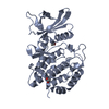
- PDB-2aqw: Structure of putative orotidine-monophosphate-decarboxylase from ... -
+ Open data

- Basic information
| Entry | Database: PDB / ID: 2aqw | ||||||
|---|---|---|---|---|---|---|---|
| Title | Structure of putative orotidine-monophosphate-decarboxylase from Plasmodium yoelii (PY01515) | ||||||
 Components Components | putative orotidine-monophosphate-decarboxylase | ||||||
 Keywords Keywords |  STRUCTURAL GENOMICS / UNKNOWN FUNCTION / decarboxylase PY01515 SGC / Structural Genomics Consortium STRUCTURAL GENOMICS / UNKNOWN FUNCTION / decarboxylase PY01515 SGC / Structural Genomics Consortium | ||||||
| Function / homology |  Function and homology informationorotidine-5'-phosphate decarboxylase / orotidine-5'-phosphate decarboxylase activity / 'de novo' UMP biosynthetic process / 'de novo' pyrimidine nucleobase biosynthetic process Function and homology informationorotidine-5'-phosphate decarboxylase / orotidine-5'-phosphate decarboxylase activity / 'de novo' UMP biosynthetic process / 'de novo' pyrimidine nucleobase biosynthetic processSimilarity search - Function | ||||||
| Biological species |  Plasmodium yoelii (eukaryote) Plasmodium yoelii (eukaryote) | ||||||
| Method | X-RAY DIFFRACTION / SYNCHROTRON / SAD / Resolution: 2 Å | ||||||
 Authors Authors | Dong, A. / Vedadi, M. / Wasney, G. / Zhao, Y. / Lew, J. / Alam, Z. / Melone, M. / Koeieradzki, I. / Edwards, A.M. / Arrowsmith, C.H. ...Dong, A. / Vedadi, M. / Wasney, G. / Zhao, Y. / Lew, J. / Alam, Z. / Melone, M. / Koeieradzki, I. / Edwards, A.M. / Arrowsmith, C.H. / Weigelt, J. / Sundstrom, M. / Bochkarev, A. / Hui, R. / Amani, M. / Structural Genomics Consortium (SGC) | ||||||
 Citation Citation | Journal: Mol.Biochem.Parasitol. / Year: 2007 Title: Genome-scale protein expression and structural biology of Plasmodium falciparum and related Apicomplexan organisms. Authors: Vedadi, M. / Lew, J. / Artz, J. / Amani, M. / Zhao, Y. / Dong, A. / Wasney, G.A. / Gao, M. / Hills, T. / Brokx, S. / Qiu, W. / Sharma, S. / Diassiti, A. / Alam, Z. / Melone, M. / Mulichak, A. ...Authors: Vedadi, M. / Lew, J. / Artz, J. / Amani, M. / Zhao, Y. / Dong, A. / Wasney, G.A. / Gao, M. / Hills, T. / Brokx, S. / Qiu, W. / Sharma, S. / Diassiti, A. / Alam, Z. / Melone, M. / Mulichak, A. / Wernimont, A. / Bray, J. / Loppnau, P. / Plotnikova, O. / Newberry, K. / Sundararajan, E. / Houston, S. / Walker, J. / Tempel, W. / Bochkarev, A. / Kozieradzki, I. / Edwards, A. / Arrowsmith, C. / Roos, D. / Kain, K. / Hui, R. | ||||||
| History |
| ||||||
| Remark 300 | Author states that biological unit for the protein is not yet known |
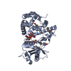
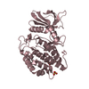
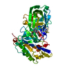
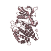
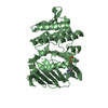
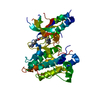
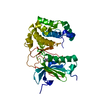
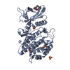
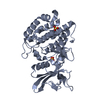
- Structure visualization
| Structure viewer | Molecule: MolmilJmol/JSmol |
|---|
- Downloads & links
-Download
| PDBx/mmCIF format | 2aqw.cif.gz | 79.6 KB | Display | PDBx/mmCIF format |
|---|---|---|---|---|
| PDB format | pdb2aqw.ent.gz | 64.4 KB | Display | PDB format |
| PDBx/mmJSON format | 2aqw.json.gz | Tree view | PDBx/mmJSON format | |
| Others |  Other downloads Other downloads |
-Validation report
| Arichive directory | https://data.pdbj.org/pub/pdb/validation_reports/aq/2aqwftp://data.pdbj.org/pub/pdb/validation_reports/aq/2aqw | HTTPS FTP |
|---|
-Related structure data
| Related structure data | 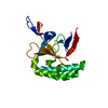 1txjC 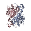 1xccC 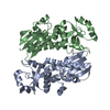 1y6zC 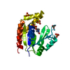 1z6gC 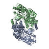 1z7dC 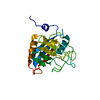 1z81C  1zo2C 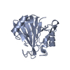 2a22C 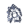 2a4aC 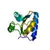 2aifC 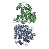 2amxC 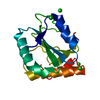 2av4C 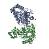 2awpC 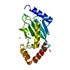 2ayvC 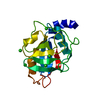 2b71C 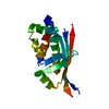 2bddC 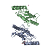 2f4zC 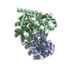 2fdsC 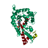 2ffcC 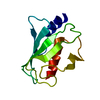 2fo3C 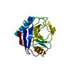 2fu0C 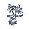 2ghiC 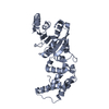 2h1rC 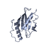 2h2yC 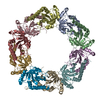 2h66C 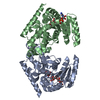 2hjrC 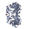 2hteC 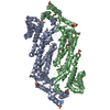 2hvgC  3pggC 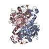 3tb2C C: citing same article ( |
|---|---|
| Similar structure data |
-Links
 PDBj
PDBj
- Assembly
| Deposited unit | 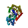
| ||||||||
|---|---|---|---|---|---|---|---|---|---|
| 1 |
| ||||||||
| 2 | 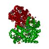
| ||||||||
| Unit cell |
| ||||||||
| Components on special symmetry positions |
|
-Components
| #1: Protein | Mass: 39291.645 Da / Num. of mol.: 1 Source method: isolated from a genetically manipulated source Source: (gene. exp.) Plasmodium yoelii (eukaryote) / Plasmid: pET28 / Production host:  Escherichia coli (E. coli) / Strain (production host): DH5-ALPHA / References: UniProt: Q7RPE4 Escherichia coli (E. coli) / Strain (production host): DH5-ALPHA / References: UniProt: Q7RPE4 | ||||
|---|---|---|---|---|---|
| #2: Chemical | ChemComp-IOD / Iodide  Mass: 126.904 Da / Num. of mol.: 6 / Source method: obtained synthetically / Formula: I Mass: 126.904 Da / Num. of mol.: 6 / Source method: obtained synthetically / Formula: I#3: Chemical | Sulfate  Mass: 96.063 Da / Num. of mol.: 3 / Source method: obtained synthetically / Formula: SO4 Mass: 96.063 Da / Num. of mol.: 3 / Source method: obtained synthetically / Formula: SO4#4: Water | ChemComp-HOH / | Water Mass: 18.015 Da / Num. of mol.: 168 / Source method: isolated from a natural source / Formula: H2O Mass: 18.015 Da / Num. of mol.: 168 / Source method: isolated from a natural source / Formula: H2O |
-Experimental details
-Experiment
| Experiment | Method: X-RAY DIFFRACTION / Number of used crystals: 2 |
|---|
- Sample preparation
| Crystal | Density Matthews: 1.9 Å3/Da / Density % sol: 35.8 % |
|---|---|
| Crystal grow | Temperature: 297 K / Method: vapor diffusion / pH: 7 Details: 2.5 M (NH4)2SO4, Bis-Tris propane pH 7.0 and 10mM NaI, VAPOR DIFFUSION, temperature 297K |
-Data collection
| Diffraction |
| ||||||||||||||||||
|---|---|---|---|---|---|---|---|---|---|---|---|---|---|---|---|---|---|---|---|
| Diffraction source |
| ||||||||||||||||||
| Detector |
| ||||||||||||||||||
| Radiation |
| ||||||||||||||||||
| Radiation wavelength |
| ||||||||||||||||||
| Reflection | Resolution: 2→20 Å / Num. all: 21954 / Num. obs: 21895 / % possible obs: 99.1 % / Observed criterion σ(F): 0 / Observed criterion σ(I): 0 / Redundancy: 6 % / Biso Wilson estimate: 28.6 Å2 / Rmerge(I) obs: 0.9 / Rsym value: 0.9 / Net I/σ(I): 9.1 | ||||||||||||||||||
| Reflection shell | Resolution: 2→2.07 Å / Redundancy: 4.8 % / Rmerge(I) obs: 0.546 / Mean I/σ(I) obs: 2.35 / Num. unique all: 2073 / Rsym value: 0.546 / % possible all: 97.1 |

- Processing
| Software |
| ||||||||||||||||||||||||||||||||||||||||||||||||||||||||||||||||||||||||||||||||||||||||||||||||||||||||||||||||||||||||||||||||||||||||||||||||||||||||||||||||||||||||||
|---|---|---|---|---|---|---|---|---|---|---|---|---|---|---|---|---|---|---|---|---|---|---|---|---|---|---|---|---|---|---|---|---|---|---|---|---|---|---|---|---|---|---|---|---|---|---|---|---|---|---|---|---|---|---|---|---|---|---|---|---|---|---|---|---|---|---|---|---|---|---|---|---|---|---|---|---|---|---|---|---|---|---|---|---|---|---|---|---|---|---|---|---|---|---|---|---|---|---|---|---|---|---|---|---|---|---|---|---|---|---|---|---|---|---|---|---|---|---|---|---|---|---|---|---|---|---|---|---|---|---|---|---|---|---|---|---|---|---|---|---|---|---|---|---|---|---|---|---|---|---|---|---|---|---|---|---|---|---|---|---|---|---|---|---|---|---|---|---|---|---|---|
| Refinement | Method to determine structure: SAD / Resolution: 2→20 Å / Cor.coef. Fo:Fc: 0.943 / Cor.coef. Fo:Fc free: 0.917 / SU B: 4.559 / SU ML: 0.131 / Cross valid method: THROUGHOUT / σ(F): 0 / σ(I): 0 / ESU R: 0.228 / ESU R Free: 0.194 / Stereochemistry target values: MAXIMUM LIKELIHOOD / Details: HYDROGENS HAVE BEEN ADDED IN THE RIDING POSITIONS
| ||||||||||||||||||||||||||||||||||||||||||||||||||||||||||||||||||||||||||||||||||||||||||||||||||||||||||||||||||||||||||||||||||||||||||||||||||||||||||||||||||||||||||
| Solvent computation | Ion probe radii: 0.8 Å / Shrinkage radii: 0.8 Å / VDW probe radii: 1.2 Å / Solvent model: MASK | ||||||||||||||||||||||||||||||||||||||||||||||||||||||||||||||||||||||||||||||||||||||||||||||||||||||||||||||||||||||||||||||||||||||||||||||||||||||||||||||||||||||||||
| Displacement parameters | Biso mean: 29.695 Å2
| ||||||||||||||||||||||||||||||||||||||||||||||||||||||||||||||||||||||||||||||||||||||||||||||||||||||||||||||||||||||||||||||||||||||||||||||||||||||||||||||||||||||||||
| Refinement step | Cycle: LAST / Resolution: 2→20 Å
| ||||||||||||||||||||||||||||||||||||||||||||||||||||||||||||||||||||||||||||||||||||||||||||||||||||||||||||||||||||||||||||||||||||||||||||||||||||||||||||||||||||||||||
| Refine LS restraints |
| ||||||||||||||||||||||||||||||||||||||||||||||||||||||||||||||||||||||||||||||||||||||||||||||||||||||||||||||||||||||||||||||||||||||||||||||||||||||||||||||||||||||||||
| LS refinement shell | Resolution: 2→2.052 Å / Total num. of bins used: 20
|
