 Movie
Movie Controller

[English] 日本語
 Yorodumi
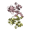
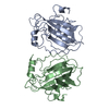
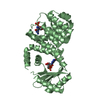
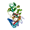
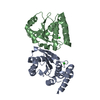
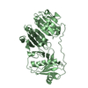
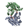
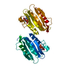
- PDB-2ae0: Crystal structure of MltA from Escherichia coli reveals a unique ... -
+ Open data

- Basic information
| Entry | Database: PDB / ID: 2ae0 | ||||||
|---|---|---|---|---|---|---|---|
| Title | Crystal structure of MltA from Escherichia coli reveals a unique lytic transglycosylase fold | ||||||
 Components Components | Membrane-bound lytic murein transglycosylase A | ||||||
 Keywords Keywords |  HYDROLASE / double-psi beta-barrel / small mixed parallel/antiparallel six stranded beta barrel / helical sub-domain HYDROLASE / double-psi beta-barrel / small mixed parallel/antiparallel six stranded beta barrel / helical sub-domain | ||||||
| Function / homology |  Function and homology information Function and homology information: / lytic transglycosylase activity / peptidoglycan turnover / hydrolase activity, hydrolyzing O-glycosyl compounds / peptidoglycan catabolic process / cell wall organization / cell outer membrane / outer membrane-bounded periplasmic space Similarity search - Function | ||||||
| Biological species |  Escherichia coli (E. coli) Escherichia coli (E. coli) | ||||||
| Method | X-RAY DIFFRACTION / SYNCHROTRON / MAD / Resolution: 2 Å | ||||||
 Authors Authors | Van Straaten, K.E. / Dijkstra, B.W. / Vollmer, W. / Thunnissen, A.M.W.H. | ||||||
 Citation Citation | Journal: J.Mol.Biol. / Year: 2005 Title: Crystal Structure of MltA from Escherichia coli Reveals a Unique Lytic Transglycosylase Fold Authors: van Straaten, K.E. / Dijkstra, B.W. / Vollmer, W. / Thunnissen, A.M.W.H. | ||||||
| History |
| ||||||
| Remark 650 | HELIX DETERMINATION METHOD: AUTHOR DETERMINED |
- Structure visualization
| Structure viewer | Molecule: MolmilJmol/JSmol |
|---|
- Downloads & links
-Download
| PDBx/mmCIF format | 2ae0.cif.gz | 84.8 KB | Display | PDBx/mmCIF format |
|---|---|---|---|---|
| PDB format | pdb2ae0.ent.gz | 63.1 KB | Display | PDB format |
| PDBx/mmJSON format | 2ae0.json.gz | Tree view | PDBx/mmJSON format | |
| Others |  Other downloads Other downloads |
-Validation report
| Arichive directory | https://data.pdbj.org/pub/pdb/validation_reports/ae/2ae0ftp://data.pdbj.org/pub/pdb/validation_reports/ae/2ae0 | HTTPS FTP |
|---|
-Related structure data
| Similar structure data |
|---|


-Links
 PDBj
PDBj


- Assembly
| Deposited unit | 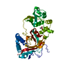
| ||||||||
|---|---|---|---|---|---|---|---|---|---|
| 1 |
| ||||||||
| Unit cell |
|
-Components
| #1: Protein | Mass: 38250.688 Da / Num. of mol.: 1 Source method: isolated from a genetically manipulated source Source: (gene. exp.) Escherichia coli (E. coli) / Gene: mlta / Production host: Escherichia coli (E. coli)References: UniProt: P0A935, Hydrolases; Glycosylases; Glycosidases, i.e. enzymes that hydrolyse O- and S-glycosyl compounds |
|---|---|
| #2: Chemical | ChemComp-EDO / Ethylene glycol  Mass: 62.068 Da / Num. of mol.: 1 / Source method: obtained synthetically / Formula: C2H6O2 Mass: 62.068 Da / Num. of mol.: 1 / Source method: obtained synthetically / Formula: C2H6O2 |
| #3: Chemical | ChemComp-ACY / Acetic acid  Mass: 60.052 Da / Num. of mol.: 1 / Source method: obtained synthetically / Formula: C2H4O2 Mass: 60.052 Da / Num. of mol.: 1 / Source method: obtained synthetically / Formula: C2H4O2 |
| #4: Water | ChemComp-HOH / Water Mass: 18.015 Da / Num. of mol.: 278 / Source method: isolated from a natural source / Formula: H2O Mass: 18.015 Da / Num. of mol.: 278 / Source method: isolated from a natural source / Formula: H2O |
-Experimental details
-Experiment
| Experiment | Method: X-RAY DIFFRACTION / Number of used crystals: 1 |
|---|
- Sample preparation
| Crystal | Density Matthews: 4.47 Å3/Da / Density % sol: 72.47 % |
|---|---|
| Crystal grow | Temperature: 280 K / Method: vapor diffusion, hanging drop / pH: 4.2 Details: PEG 8000, sodium chloride, sodium acetate, pH 4.2, VAPOR DIFFUSION, HANGING DROP, temperature 280K |
-Data collection
| Diffraction | Mean temperature: 100 K | |||||||||||||||
|---|---|---|---|---|---|---|---|---|---|---|---|---|---|---|---|---|
| Diffraction source | Source: SYNCHROTRON / Site: ESRF  / Beamline: ID14-1 / Wavelength: 0.934, 0.9794, 0.9792, 0.9393 / Beamline: ID14-1 / Wavelength: 0.934, 0.9794, 0.9792, 0.9393 | |||||||||||||||
| Detector | Type: ADSC QUANTUM 4 / Detector: CCD / Date: Sep 23, 2002 | |||||||||||||||
| Radiation | Monochromator: diamonds (111), Ge(220) / Protocol: MAD / Monochromatic (M) / Laue (L): M / Scattering type: x-ray | |||||||||||||||
| Radiation wavelength |
| |||||||||||||||
| Reflection | Resolution: 2→30 Å / Num. all: 46854 / Num. obs: 46854 / % possible obs: 100 % / Observed criterion σ(I): 2 / Rmerge(I) obs: 0.049 / Χ2: 0.934 | |||||||||||||||
| Reflection shell | Resolution: 2→2.07 Å / % possible obs: 100 % / Rmerge(I) obs: 0.501 / Num. measured obs: 4652 / Χ2: 0.866 / % possible all: 100 |
- Processing
| Software |
| ||||||||||||||||||||||||||||||||||||||||||||||||||||||||||||||||||||||
|---|---|---|---|---|---|---|---|---|---|---|---|---|---|---|---|---|---|---|---|---|---|---|---|---|---|---|---|---|---|---|---|---|---|---|---|---|---|---|---|---|---|---|---|---|---|---|---|---|---|---|---|---|---|---|---|---|---|---|---|---|---|---|---|---|---|---|---|---|---|---|---|
| Refinement | Method to determine structure: MAD / Resolution: 2→30 Å / Cor.coef. Fo:Fc: 0.953 / Cor.coef. Fo:Fc free: 0.932 / SU B: 3.13 / SU ML: 0.086 / TLS residual ADP flag: LIKELY RESIDUAL / Isotropic thermal model: isotropic / Cross valid method: THROUGHOUT / σ(F): 0 / ESU R: 0.124 / ESU R Free: 0.124 / Stereochemistry target values: MAXIMUM LIKELIHOOD
| ||||||||||||||||||||||||||||||||||||||||||||||||||||||||||||||||||||||
| Solvent computation | Ion probe radii: 0.8 Å / Shrinkage radii: 0.8 Å / VDW probe radii: 1.4 Å / Solvent model: BABINET MODEL WITH MASK | ||||||||||||||||||||||||||||||||||||||||||||||||||||||||||||||||||||||
| Displacement parameters | Biso mean: 37.227 Å2
| ||||||||||||||||||||||||||||||||||||||||||||||||||||||||||||||||||||||
| Refinement step | Cycle: LAST / Resolution: 2→30 Å
| ||||||||||||||||||||||||||||||||||||||||||||||||||||||||||||||||||||||
| Refine LS restraints |
| ||||||||||||||||||||||||||||||||||||||||||||||||||||||||||||||||||||||
| LS refinement shell | Resolution: 2→2.02 Å / Total num. of bins used: 50
| ||||||||||||||||||||||||||||||||||||||||||||||||||||||||||||||||||||||
| Refinement TLS params. | Method: refined / Origin x: 3.796 Å / Origin y: 48.52 Å / Origin z: 52.515 Å
|
