 Movie
Movie Controller

[English] 日本語
 Yorodumi
- PDB-2aac: ESCHERCHIA COLI GENE REGULATORY PROTEIN ARAC COMPLEXED WITH D-FUCOSE -
+ Open data

- Basic information
| Entry | Database: PDB / ID: 2aac | ||||||
|---|---|---|---|---|---|---|---|
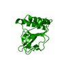
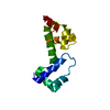
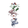
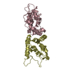
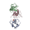
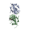
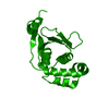
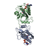
| Title | ESCHERCHIA COLI GENE REGULATORY PROTEIN ARAC COMPLEXED WITH D-FUCOSE | ||||||
 Components Components | ARAC | ||||||
 Keywords Keywords |  TRANSCRIPTION FACTOR / CARBOHYDRATE BINDING / COILED-COIL / JELLY ROLL TRANSCRIPTION FACTOR / CARBOHYDRATE BINDING / COILED-COIL / JELLY ROLL | ||||||
| Function / homology |  Function and homology information Function and homology informationarabinose catabolic process / DNA-binding transcription repressor activity / protein-DNA complex / transcription cis-regulatory region binding / identical protein binding / cytosolSimilarity search - Function | ||||||
| Biological species |  Escherichia coli (E. coli) Escherichia coli (E. coli) | ||||||
| Method | X-RAY DIFFRACTION / SYNCHROTRON / RIGID-BODY REFINEMENT, DIFFERENCE FOURIER MAPS / Resolution: 1.6 Å | ||||||
 Authors Authors | Soisson, S.M. / Wolberger, C. | ||||||
 Citation Citation | Journal: J.Mol.Biol. / Year: 1997 Title: The 1.6 A crystal structure of the AraC sugar-binding and dimerization domain complexed with D-fucose. Authors: Soisson, S.M. / MacDougall-Shackleton, B. / Schleif, R. / Wolberger, C. | ||||||
| History |
|
- Structure visualization
| Structure viewer | Molecule: MolmilJmol/JSmol |
|---|
- Downloads & links
-Download
| PDBx/mmCIF format | 2aac.cif.gz | 83.7 KB | Display | PDBx/mmCIF format |
|---|---|---|---|---|
| PDB format | pdb2aac.ent.gz | 65 KB | Display | PDB format |
| PDBx/mmJSON format | 2aac.json.gz | Tree view | PDBx/mmJSON format | |
| Others |  Other downloads Other downloads |
-Validation report
| Arichive directory | https://data.pdbj.org/pub/pdb/validation_reports/aa/2aacftp://data.pdbj.org/pub/pdb/validation_reports/aa/2aac | HTTPS FTP |
|---|
-Related structure data
| Related structure data |  2arcS S: Starting model for refinement |
|---|---|
| Similar structure data |


-Links
 PDBj
PDBj

- Assembly
| Deposited unit | 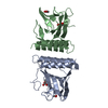
| ||||||||
|---|---|---|---|---|---|---|---|---|---|
| 1 |
| ||||||||
| Unit cell |
| ||||||||
| Noncrystallographic symmetry (NCS) | NCS oper: (Code: given Matrix: (-0.191475, 0.4722, -0.860444), Vector : |
-Components
| #1: Protein | Mass: 20547.184 Da / Num. of mol.: 2 / Fragment: SUGAR-BINDING/DIMERIZATION Source method: isolated from a genetically manipulated source Source: (gene. exp.) Escherichia coli (E. coli) / Gene: ARAC / Gene (production host): ARAC / Production host: Escherichia coli (E. coli) / References: UniProt: P0A9E0#2: Sugar | Fucose  Type: D-saccharide, beta linking / Mass: 164.156 Da / Num. of mol.: 2 Type: D-saccharide, beta linking / Mass: 164.156 Da / Num. of mol.: 2Source method: isolated from a genetically manipulated source Formula: C6H12O5 #3: Chemical | Acetic acid  Mass: 60.052 Da / Num. of mol.: 2 / Source method: obtained synthetically / Formula: C2H4O2 Mass: 60.052 Da / Num. of mol.: 2 / Source method: obtained synthetically / Formula: C2H4O2#4: Water | ChemComp-HOH / | Water Mass: 18.015 Da / Num. of mol.: 405 / Source method: isolated from a natural source / Formula: H2O Mass: 18.015 Da / Num. of mol.: 405 / Source method: isolated from a natural source / Formula: H2O |
|---|
-Experimental details
-Experiment
| Experiment | Method: X-RAY DIFFRACTION / Number of used crystals: 1 |
|---|
- Sample preparation
| Crystal | Density Matthews: 2.23 Å3/Da / Density % sol: 45 % | ||||||||||||||||||||
|---|---|---|---|---|---|---|---|---|---|---|---|---|---|---|---|---|---|---|---|---|---|
| Crystal grow | pH: 5.5 Details: PROTEIN WAS CRYSTALLIZED BY MICROSEEDING FROM 24% PEG 4000, 100 MM SODIUM CITRATE PH 5.5 200 MM AMMONIUM ACETATE | ||||||||||||||||||||
| Crystal grow | *PLUS pH: 7.25 / Method: vapor diffusion, hanging dropDetails: used to seeding, Soisson, S.M., (1997) Science, 276, 421. | ||||||||||||||||||||
| Components of the solutions | *PLUS
|
-Data collection
| Diffraction | Mean temperature: 100 K |
|---|---|
| Diffraction source | Source: SYNCHROTRON / Site: NSLS  / Beamline: X4A / Wavelength: 0.689 / Beamline: X4A / Wavelength: 0.689 |
| Detector | Type: FUJI / Detector: IMAGE PLATE / Date: Sep 1, 1995 / Details: SPHERICAL RH COATED MIRROR |
| Radiation | Monochromator: SAGITALLY FOCUSED SI(111) DOUBLE CRYSTAL MONOCHROMATOR Monochromatic (M) / Laue (L): M / Scattering type: x-ray |
| Radiation wavelength | Wavelength: 0.689 Å / Relative weight: 1 |
| Reflection | Resolution: 1.6→30 Å / Num. obs: 47363 / % possible obs: 99.9 % / Observed criterion σ(I): 2 / Redundancy: 5.2 % / Biso Wilson estimate: 12.31 Å2 / Rmerge(I) obs: 0.08 / Rsym value: 0.08 / Net I/σ(I): 9.7 |
| Reflection shell | Resolution: 1.6→1.67 Å / Redundancy: 3.4 % / Rmerge(I) obs: 0.08 / Mean I/σ(I) obs: 2.5 / Rsym value: 0.39 / % possible all: 99 |
| Reflection | *PLUS Num. obs: 57378 / Num. measured all: 293876 / Rmerge(I) obs: 0.09 |
- Processing
| Software |
| ||||||||||||||||||||||||||||||||||||||||||||||||||||||||||||||||||||||||||||||||
|---|---|---|---|---|---|---|---|---|---|---|---|---|---|---|---|---|---|---|---|---|---|---|---|---|---|---|---|---|---|---|---|---|---|---|---|---|---|---|---|---|---|---|---|---|---|---|---|---|---|---|---|---|---|---|---|---|---|---|---|---|---|---|---|---|---|---|---|---|---|---|---|---|---|---|---|---|---|---|---|---|---|
| Refinement | Method to determine structure: RIGID-BODY REFINEMENT, DIFFERENCE FOURIER MAPS Starting model: PDB ENTRY 2ARC Resolution: 1.6→7 Å / Rfactor Rfree error: 0.003 / Data cutoff high absF: 100000 / Data cutoff low absF: 0.01 / Isotropic thermal model: RESTRAINED / Cross valid method: THROUGHOUT / σ(F): 2
| ||||||||||||||||||||||||||||||||||||||||||||||||||||||||||||||||||||||||||||||||
| Displacement parameters | Biso mean: 12.48 Å2
| ||||||||||||||||||||||||||||||||||||||||||||||||||||||||||||||||||||||||||||||||
| Refine analyze | Luzzati coordinate error obs: 0.15 Å / Luzzati d res low obs: 30 Å | ||||||||||||||||||||||||||||||||||||||||||||||||||||||||||||||||||||||||||||||||
| Refinement step | Cycle: LAST / Resolution: 1.6→7 Å
| ||||||||||||||||||||||||||||||||||||||||||||||||||||||||||||||||||||||||||||||||
| Refine LS restraints |
| ||||||||||||||||||||||||||||||||||||||||||||||||||||||||||||||||||||||||||||||||
| LS refinement shell | Resolution: 1.6→1.67 Å / Rfactor Rfree error: 0.012 / Total num. of bins used: 8
| ||||||||||||||||||||||||||||||||||||||||||||||||||||||||||||||||||||||||||||||||
| Xplor file |
| ||||||||||||||||||||||||||||||||||||||||||||||||||||||||||||||||||||||||||||||||
| Software | *PLUS Name: X-PLOR / Version: 3.8 / Classification: refinement | ||||||||||||||||||||||||||||||||||||||||||||||||||||||||||||||||||||||||||||||||
| Refine LS restraints | *PLUS
|
